Scene 1 (0s)
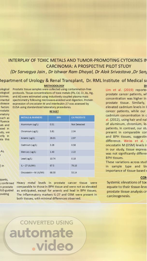
INTERPLAY OF TOXIC METALS AND TUMOR-PROMOTING CYTOKINES CARCINOMA: A PROSPECTIVE PILOT STUDY (Dr Sarvagya Jain , Dr Ishwar Ram Dhayal, Dr Alok Srivastava ,Dr Sanj )epartment of Urology & Renal Transplant, Dr. RML Institute of Medical sc -cal ological tcomes. disease factus rostatic •rnatory such as fLence als and in in ite this As, •ants, METH ODOLOGY Prostatic tisue samples conected ccntaminatim•free protocoÉ. Tissue concentrations of tokk metals (Pb, Cd, Cr, As, kg. and A1) were using inductively plasma mass spectronz•try following microwaveassisted aca digestion. Protein expressim of oncostatin M and interkukin-27 was assessai by ELISA LSing prxedures. RESULT AIL" (ug/L) Arsenic Cadm ium Lead ( IL -27 confirnEd n prostatic Ovid Heavy metal levels in prostate cancer tissue were comparable to those in BPH tissue and were not as elevated as anticipated, except for arsenic and lead in BPH tissues. The inflammatory markers IL-27 and OSM were preænt in both tissues, with minimal differences observed. Lim et al. (2019) reporte. prostate cancer patients, concentration was higher in prostate tissue. Similarly, elevated cadmium levels in cancer patients, while our cadmium concentration in c of aluminum, chromium, le, patients. In contrast, our sti present in comparable con and BPH tissues, suggestin difference. Weiss et al. oncostatin M (OSMI levels ir in our study, tissue express was not significantly differer BPH tissues. These variations across stud in sample type and bit importance of tissue-based Systemic elevations Of tox equate to their tissue-leve prostate tissue analysis carcinogenesis..