Scene 1 (0s)
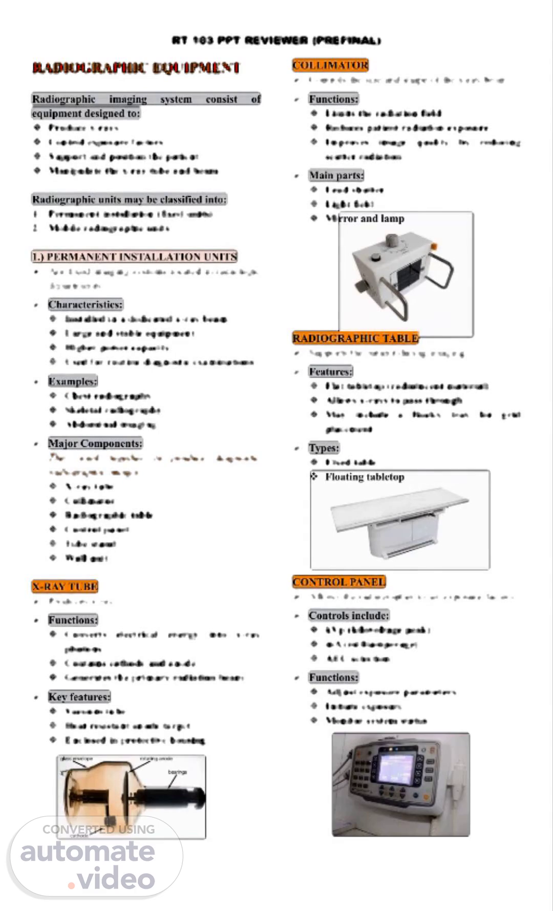
RT 103 PPT REVIEWER (PREFINAL) RADIOGRAPHIC EQUIPMENT Radiographic imaging system consist of equipment designed to: ❖ Produce x-rays ❖ Control exposure factors ❖ Support and position the patient ❖ Manipulate the x-ray tube and beam Radiographic units may be classified into: 1. Permanent installation (fixed units) 2. Mobile radiographic units 1.) PERMANENT INSTALLATION UNITS ➢ Are fixed imaging systems located in radiologic departments. ➢ Characteristics: ❖ Installed in a dedicated x-ray beam ❖ Large and stable equipment ❖ Higher power capacity ❖ Used for routine diagnostic examinations ➢ Examples: ❖ Chest radiography ❖ Skeletal radiography ❖ Abdominal imaging ➢ Major Components: They work together to produce diagnostic radiographic images ❖ X-ray tube ❖ Collimator ❖ Radiographic table ❖ Control panel ❖ Tube stand ❖ Wall unit X-RAY TUBE ➢ Produces x-ray. ➢ Functions: ❖ Converts electrical energy into x-ray photons ❖ Contains cathode and anode ❖ Generates the primary radiation beam ➢ Key features: ❖ Vacuum tube ❖ Heat resistant anode target ❖ Enclosed in protective housing COLLIMATOR ➢ Controls the size and shape of the x-ray beam. ➢ Functions: ❖ Limits the radiation field ❖ Reduces patient radiation exposure ❖ Improves image quality by reducing scatter radiation ➢ Main parts: ❖ Lead shutter ❖ Light field ❖ Mirror and lamp RADIOGRAPHIC TABLE ➢ Supports the patient during imaging. ➢ Features: ❖ Flat tabletop (radiolucent material) ❖ Allows x-rays to pass through ❖ May include a Bucky tray for grid placement ➢ Types: ❖ Fixed table ❖ Floating tabletop CONTROL PANEL ➢ Allows the radiographer to set exposure factors. ➢ Controls include: ❖ kVp (kilovoltage peak) ❖ mA (milliamperage) ❖ AEC selection ➢ Functions: ❖ Adjust exposure parameters ❖ Initiate exposure ❖ Monitor system status.
Scene 2 (1m 48s)
[Audio] The major components of radiographic equipment are the tube stand, wall unit/ upright bucky, and x-ray tube. These components work together to support and enable movement of the x-ray tube, allowing accurate positioning of the x-ray beam and alignment with the patient and image receptor. The x-ray tube produces the x-ray beam through conversion of electromagnetic energy, contained within a protective housing. The tube stand supports vertical, horizontal, rotation, and other movements of the x-ray tube, while the wall unit/upright bucky is used for standing radiographic examinations. The digital detector system and power supply also play crucial roles in producing high-quality radiographic images..
Scene 3 (2m 33s)
[Audio] The floor-to-ceiling support system consists of a single column with rollers at each end. This design allows the x-ray tube to slide up and down the column as the column rotates. The window on the enclosure enables maximum emission of x-rays while minimizing absorption and scatter of photons. The vacuum within the enclosure ensures that electrons flow freely from the cathode to the anode without encountering air molecules, thereby increasing the efficiency of x-ray production and reducing heat generation. Additionally, this setup reduces the risk of tube arcing. The enclosure itself maintains a high vacuum pressure below 10^-5 mmHg, ensuring optimal conditions for the x-ray tube's operation. Furthermore, the use of a protective housing, such as a lead-lined metal container, provides additional protection against radiation leaks and helps maintain electric constant potential between the electrons and the enclosure. The internal components, including the cathode and anode assemblies, are carefully designed to withstand thermal and mechanical stresses, with features like thermal cushions and insulators to prevent damage. Overall, these components work together to ensure accurate and reliable x-ray imaging..
Scene 4 (3m 55s)
[Audio] The x-ray tube consists of two main parts: the cathode and the anode. The cathode is the negative side of the x-ray tube and produces a thermionic cloud, which helps focus the electron stream. The most common materials used for the anode are copper, molybdenum, and graphite. The primary parts of the x-ray tube include the filament and focusing cup. The filament is a coil of wire that emits electrons when heated, providing sufficient resistance to the flow of electrons. The focusing cup is a metal shroud that surrounds the filament and is designed to house it. The focusing cup also serves to narrow the thermionic cloud. The anode is a positively charged electrode that conducts electricity, radiates heat, and contains a target. There are two types of anodes: stationary and rotating. The stationary anode has a static target area and is made of rhodium-alloyed tungsten, while the rotating anode is made of tungsten and provides higher thermionic emission. The anode plays a crucial role in producing x-rays and is essential for the proper functioning of the x-ray tube..
Scene 5 (5m 9s)
[Audio] The rotating anode is used in X-ray tubes to increase the interaction area between the electron beam and the target material. This increases the efficiency of X-ray production. The most common rotational speed for rotating anodes is between 3200-3600 revolutions per minute. A large focal spot is associated with the large filament and is used for imaging large body parts, such as the head or torso. It also allows for the tolerance of high heat loading and is suitable for heavy tube loads. However, it has some disadvantages, including focal spot blooming. High-capacity X-ray tubes operate at speeds above 10000 revolutions per minute, resulting in increased Ma and OID values. These values are caused by increased electron beam energy and reduced scattering. Faster rotations and better heat dissipation are possible with high tube currents and lower exposure times. The electromagnetic induction motor powers these fast rotations. The target is the stationary area where the electrons strike the anode, typically made of tungsten alloy embedded in the copper anode. The high atomic number of the target results in high efficiency X-ray production and high-energy X-rays. The high thermal conductivity of the anode enables efficient heat dissipation. The rotating anode is powered by an electromagnetic induction motor, which provides faster rotations and better heat dissipation. The anode heel effect occurs due to the line focus principle, causing absorption of X-rays in the heel of the target. This effect changes the effective FSS and FATCAT values. The normal operating temperature of the focal track is around 1000-2000°C. The focal spot is the region on the anode target where electrons interact..