Scene 2 (5s)
SUMMARY OF PROJECTIONS PROJECTIONS, POSITIONS. & METHODS Page 260 267 268 270 Essential Anatomy Hysterosalpingography AbdOmen: Abdomen: pelvimetry Seminal ducts Projection AP lateral axid. oblique Laterd AP or AP oblique Position Method COLCHER-SUSSMAN COLCHER-SUSSMAN Icons in the Essential column indicate projections frequently performed in the United States and Canada. Stucients should be competent in these projections.
Scene 4 (25s)
Tætjcular ater•y• deferens Head Of epiddyrnis Testis Fig. 19-7 Frontd section of testes cnd ductus deferens. Sacrum Prostate Urethra 19-8 section showhg male genital systern..
Scene 5 (35s)
SUMMARY OF ANATOMY• Female reproductive systern ovaries uterine tubes uterus vagha Ovaries ova rnesovarium medulla cortex ovarian follicles gaafian follicle ovulatiorl Uterine tubes (Fallopian fthes) isthmus ampulla infundibulum fimbriae cilia Uterus fundus body isthrnus cervlx uterine ostium endometrium Vagina mucosa vaginal vestibule vaginal orifice urethral orifice Fetd development zygote embryo fetus placenta Male reproductive system testes ductus deferens (vas deferens) prostate ejaculatory ducts seminal vesicles bulbourethral glands penis scrotum Testes epididymis Ductus deferens ampulla.
Scene 6 (44s)
SUMMARY OF PATHOLOGY Condition Adhesion Endometrial Polyp Fallopian Tube Obstruction Fistda Tumor Dermoid Cyst Uterhe Fbroid Definition Union of two surfaces that are normally separate Growth or rnass gotruding from the endometrium Condition preventng normat flow through the fallopian tl-öe Abnormal connection between two internd orgms or between an organ and tre body surface New tissue gowth where cell proifgation is uncontroled Tumor of the ovary filled with sebaceous material and hair Smooth-muscle tumor of the uterus.
Scene 7 (1m 2s)
Female Radiography NONPREGNANT PATIENT. Hysterosalpingography (HSG) is performed by a physician with spot radiographs made while the patient is in the supine position on a fluoroscopic table. The examination may also be performed by the physician with conventional radiographs obtained using an overhead tube. When fluoroscopy is used, spot radiographs may be the only i mages obtained. Hysterosalpingography involves the introduction of a radiopaque contrast medium through a uterine cannula. The procedure is performed to determine the size, shape, and position of the uterus and uterine tubes; to delineate lesions such as polyps, submucous tumor masses, or fistulous tracts; and to investigate the patency of the uterine tubes in patients who have been unable to conceive.
Scene 9 (1m 38s)
V aginog r ap h y i s p e r f orm e d t o i n v e s t i g a t e congenital abnormalities, vaginal fistulae, and other pathologic conditions involving the vagina. Contrast media Various opaque media are used in examinations of the female genital passages. The water- soluble contrast media employed for intravenous urography are widely used for hysterosalpingography and vaginography..
Scene 10 (2m 1s)
Fetography is the demonstration of the fetus in utero. If possible, this examination technique is avoided until after the eighteenth week of gestation because of the danger of radiation-induced fetal malformations. Fetography is employed to detect su pected abnormalities of development, to confirm su pected fetal death, to determine the presentation and position of the fetus, and to determine whether the pregnancy is single or multiple..
Scene 11 (2m 26s)
Radiographic pelvimetry and cephalometry Most pelvimetry techniques have been replaced by sonography. Thus the Ball and Thoms methods have been deleted from this edition. (See the eventh or earlier edition of this atlas for descriptions of these methods.) However, obtainable pelvic measurements and the ColcherSussman method of pelvimetry are described here..
Scene 12 (2m 59s)
Pelvimetry AP PROJECTION COLCHEP-SLISSMAN METHOD The two projections (AP and lateral) em- ployed in this method Of re- quire the use of the Colcher-Sussman pelvi meter. This device consists of a metal ruler perfcyated at centimeter intervals and mounted on a small stand in such a way that it is always parallel to the plane Of the IR. The ruler can be rotated in a complete circle and adjusted for height (Fig. 19-25). Image receptor: 35 x 43 cm for each exposure Position Of patient Place the patient in the supine position, and center the midsagittal plane of the body to the midline Of the grid. Position of part Flex the patient •s knees to elevate the forepelvis. and separate the thighs enough to permit correct placement Of the Center the horizontal ruler to the gluteal fold at the level of the ischial tuberosities. The tuberosities are easily palpated through the median part Of the buttocks. If preferred, local ize the tuberosities by placing the ruler 10 cm below the superior border Of the pubic symphysis (Fig. 19-26). Center the IR 1 inches (3.8 cm) supe— rior to the pubic symphysis (Fig. 19-27). Fig. 19-25 Colcher-Susyncn roer. Fig. 19-26 Pélvrnetry. AP projection. with ruler place. E Y-get T — Outset.
Scene 13 (3m 46s)
palpated through the median part of the buttocks. If preferred, localize the tuberosities by plæing the ruler 10 cm below the superior border of the pubic symphysis (Fig. 19-26). • Center the IR 1 inches (3.8 cm) supe- rior to the pubic symphysis (Fig. 19-27). • Respiration: After determining that the fetus is quiet. instruct the patient to respiration at the end of expiration. Central ray • Perpendicular to the midpoint of the IR and I i/2 inches (3.8 cm) superior to the pubic symphysis EVALUATION CRITERIA The following should be clearly demon- strated: • Entire pelvis • Metal ruler with centimeter markings visible • Density permitting visualization of all pelvic landmarks and intersecting di- ameters • No rotation of the }rlvis • Entire fetal head E Wet - Midpetvqs T - Outlet Pubic Ichial Fig 19-27 Pelvirnetry. AP grojection..
Scene 14 (4m 18s)
Pelvimetry LATERAL PROJECTION COLCHER-SUSSMAN METHOD R or L position Image receptor: 35 x 43 cm Position of patient Ask the patient to turn to a lateral posi- tion. and center the midcoronal plane of the patient's body to the midline of the table. Position of part • Partially extend the patient's thighs so that they do not obscure the pubic bones. • Place sandbags under and between the patient's knees and ankles to immobi- lize the legs. • Place a folded sheet or other suitable support under the lower thorax. and ad- just the support so that the long axis of the lumbar vertebrae is parallel with the tabletop. Adjust the patient's body in a true lat- • eral position. Turn the ruler lengthwise, and adjust its • height to coincide with the midsagittal plane of the patient's body. • Place the pelvimeter so that the metal ruler lies within the upper part of the gluteal fold and against the midsacrum (Fig. 19-28). Center the IR at the level of the most • prominent point of the greater trochanter (Fig. 19-29). Respiration: Suspend at the end of ex- • piration. Central ray • PerErndicuIar to the most prominent point of the greater trochanter EVALUATION CRITERIA The following should be clearly demon- strated: Superimposed hips and femora No superimposition of the pubic sym- physis by the femurs Entire pelvis, sacrum, and coccyx Metal ruler with centimeter markings visible Ihnsity permitting visualization of all pelvic landmarks and intersecting diameters Entire fetal head Fig. 19-28 Petvirnetrv. laterd projection with ruler in plcye..
Scene 15 (5m 11s)
LOCALIZATION OF INTRAUTERINE DEVICES Intrauterine devices (IUDs) remain a contraceptive option. Occasionally an IUD becomes dislocated from the uterine cavity. If this occurs, the exact location of the device must be determined, in some cases by radiography. Therefore it is necessary to become acquainted with the radiographic appearance of IUD . The physician first performs a pelvic examination to determine the location of the IUD. If the IUD is not located, the physician passes a sterile probe into the uterine cavity and radiographs are taken. AP and lateral projections of the abdomen are suggested for IUD localization. Occasionally, oblique projections are indicated. Most IUDs are radiopaque because of their inherent metallic density or because of barium impregnated in the plastic during their manufacture. It should be emphasized that radiography alone is not a reliable way to diagnose extrauterine localization of an IUD..
Scene 16 (5m 47s)
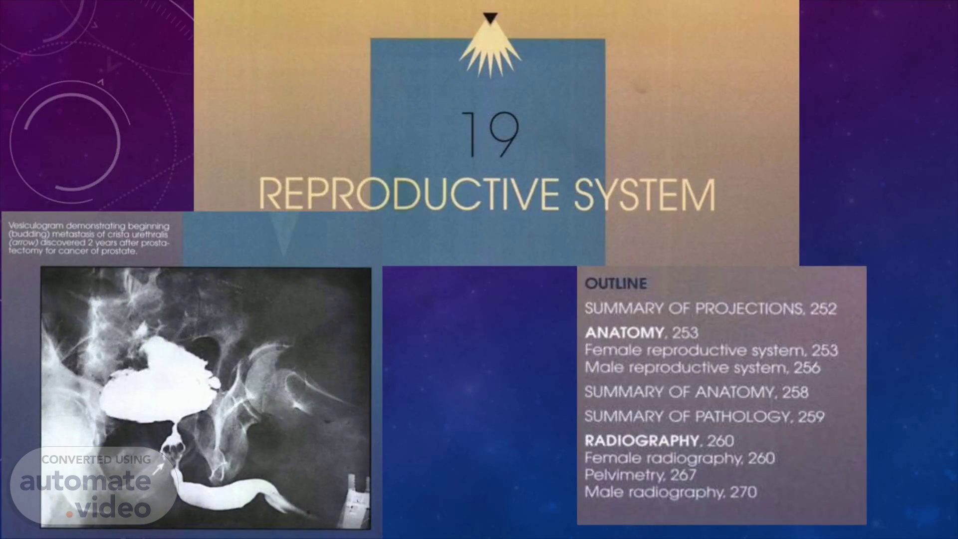
Male Radiography SEMINAL DUCTS. Radiologic examinations of the seminal ducts'·3 are performed in the investigation of selected genitourinary abnormalities such as cysts, abscesses, tumors, inflammation, and sterility. The regional terms applied to these examinations are vesiculography, epididymography, and, when combined, epididymovesiculography. The contrast medium employed for these procedures is one of the water-soluble, iodinated compounds used for intravenous urography. A gaseous contrast medium can be injected into each scrotal ac to improve contrast in the examination of extra pelvic structures..
Scene 17 (6m 12s)
The seminal vesicle are sometimes opacified directly by urethroscopic catheterization of the ejaculatory ducts. More frequently the entire duct system is inspected by introducing contrast solution into the canals by way of the ductus deferens. This requires small bi lateral incisions in the upper part of the scrotum for the exposure and identification of these ducts. The needle that is used to inject the contrast medium is inserted into the duct in the direction of the portion of the tract under investigation-distally for study of the extra pelvic ducts and then proximally for study of the intrapelvic ducts..
Scene 18 (6m 38s)
P R O S TA T E. Prostatography is a term applied to the investigation of the prostate by radiographic, cystographic, or vesiculographic procedures. It is seldom performed today because of advancements in the diagnostic value of ultrasonography. Radiographic examination of the prostate gland was described in the eighth and earlier editions of this atlas..
Scene 20 (7m 3s)
Respiratory System. 4.1. Indications and contraindications 4.2. Preparation of patient 4.3. Contrast medium 4.4. Type of examination 4.4.1. Bronchography 4.5. Modifications of positioning.
Scene 22 (7m 22s)
• Bronchography is a special study of the bronchial tree by means of introduction of a contrast medium into a particular bronchus usually under fluoroscopic control. The contrast media are nonirritating and sufficiently radiopaque to allow good visualization of the bronchi. After the radiographic examination is completed, the patient is asked to cough and expectorate the contrast medium. Bronchography Posteroanterior Bronchogram.
Scene 23 (7m 37s)
Bronchography o Until recently bronchography was the definitive investigation for the diagnosis of bronchiectasis and for assessing the extent of the disease. o High-resolution CT is now widely preferred. Other indications: o Recurrent haemoptysis when all other investigations are negative. o Bronchopleural fistulas. o Congenital lesions like sequestration and agenesis. Contraindications: 0 Severe or partial impairment of pulmonary functions. Massive haemoptysis Resent pneumonia Active TB History of allergy..
Scene 24 (7m 55s)
PURPOSE To detect bronchial obstruction such as foreign bodies and tumors Indications: bronchial obstructions (e.g. foreign bodies, tumors, cysts or cavities, bronchiectasis) CLIENT PREPARATION • Obtain a signed consent form. Check that the consent form is signed premeditation is given • Explain the procedure of the test. Gradually clients are extremely apprehensive about this test and are fearful that they may be unable to breath • Reassure the client that airway will not be blocked. Inform the patient that he or she may have a sore throat after the test as the result of catheter irritation • Obtain history of hypersensitivity to anesthetics, iodine and X-ray dyes. Usually the client will receive an expectorant several days before the test to loose secretions • Record the vital signs.
Scene 25 (8m 25s)
PROCEDURE • A consent form should be signed • The client should be NPO for 6 to 8 hours before the test • Oral hygiene should be given the night before the test and in the morning. This will decrease the number of bacteria that could be introduced into the lungs • Postural drainage is performed for 3 days before the test. This procedure aids in the removal of bronchial mucus and secretions • A sedative and atropine are usually given 1 hour before the tests. The sedative/tranquilizer is to promote relaxation; atropine is to reduce secretions during the test • A topical anesthetic is sprayed into the pharynx and trachea. A catheter is passed through the nose into the trachea, and a local anesthetic and iodized contrast liquid are injected through the catheter • The client is usually asked to change body positions so that the contrast dye can reach most areas of the bronchial tree • Following the bronchography procedure, the client may receive nebulization and should perform postural drainage to remove contrast dye. Food and fluids are restricted until the gag (cough) reflex is present.
Scene 26 (9m 5s)
POST-PROCEDURAL CARE • Assess for signs and symptoms of laryngeal edema (e.g. dyspnea, hoarseness, apprehension). This could be caused by a traumatic insertion of the catheter • Assess for allergic reaction to the anesthetic and iodized contrast dye (e.g. apprehension, flushing, rash, urticaria, dyspnea, tachycardia and hypotension) • Check the gag reflex to see that it has returned before offering food and fluids. Have the client swallow and cough or tickle the posterior pharynx will a cotton swab; if gag reflex is present, offer ice chips or sips of water before food • Monitor vital signs. The temperature may be slightly elevated for 1 or 2 days after the test • Checks breathe signs. If bronchi and fever are present, notify the health care providers and record on the client's chart • Have the client perform postural drainage post-test? This procedure helps with the removal of the contrast dye. Physiologic damage will not occur if some of the dye remains in the lungs for a period of time • Offer throat lozenges or an ordered medication for answer their questions • Be supportive of the client and family. Be available to answer their questions.
Scene 27 (9m 50s)
Contrast Media Contrast media used was dianosil 20ml for unilateral and 40ml for bilateral, as it was oily based low viscosity iodinated contrast media which gives better visualization. Now a days non-ionic monomer (iohexol) contrast media use for bronchography. Bronchografin - Mixer of lignocaine, carboxymethyl cellulose, and conray 280..
Scene 28 (10m 5s)
Technique • The injection of the contrast medium should be done under fluoroscopic guidance using the spot film device for recording the bronchial anatomy in the frontal, oblique and lateral projection. • Average of 15-20 ml of contrast. • The patient is placed in reverse Trendelenburg position, which allows the CM flow into the bronchus by gravity. • Different positions and variable quantity of contrast are required to fill the segments of upper lobe, middle lobe and lower lobe..
Scene 29 (10m 26s)
Right side Examination: Liftthe left side to 30 degree, so contrast flows in the upper, middle and lower lobes of the lung , with the help of gravity. > Left side Examination: Lift the right side to 30 degree so the contrast media flows in the upper and lower lobe of the lung, with the help of gravity. The patient must be instructed for shallow breathing and avoid coughing during the procedure, during injection of CM under fluoroscopy. Use suction machine for suction of saliva during the procedure..
Scene 30 (10m 50s)
>Post tussive or delayed Bronchogram- A segment which appeared dilated and incompletely filled may reveal good filling after vigorous coughing to exclude bronchiectasis. Firm thumping over the non-filling segment helps to suck the contrast into the peripheral bronchi, especially in situations of segmented or lobar atelectasis where the lobe cannot expand and hence suction effect into the bronchus is inadequate. After completion of the examination, the catheter or bronchoscope will be removed and the patient is shifted in the observation room..
Scene 31 (11m 11s)
After Care: • Keep the patient under observation blood pressure, heart rate, oxygen level, fluid balance and other vital sign must be monitored. • Observe for respiratory dysfunction; give oxygen if necessary. • Instruct the patient for coughing, to the split the remaining contrast media from airways. • After the 24 hours of procedure, X-ray PA Chest performs to assess the remaining contrast media in airways. • Postural drainage, antibiotic cover, plenty of oral fluids..
Scene 32 (11m 32s)
END OF PRESENTATION T H ANK Y OU F O R Y OUR A T T EN T ION..