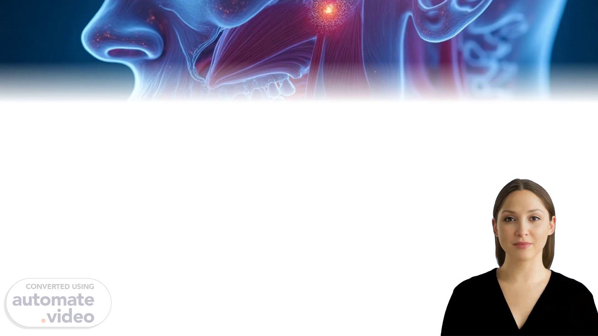
Scene 1 (0s)
[Virtual Presenter] The muscles of mastication are primarily located in the face and neck region. They include the masseter muscle, the temporalis muscle, the medial pterygoid muscle, the lateral pterygoid muscle, and the digastric muscle. Each of these muscles has distinct functions that contribute to the overall movement of the jaw. The masseter muscle is one of the most prominent muscles of mastication. Its primary function is to elevate the mandible, which means it helps to close the jaw. The temporalis muscle also plays a significant role in elevating the mandible. However, its secondary function is to rotate the mandible laterally, which allows for more efficient chewing. The medial pterygoid muscle assists in depressing the mandible, which enables the opening of the mouth. This action is essential for various activities such as eating, speaking, and yawning. The lateral pterygoid muscle is responsible for rotating the mandible medially, which facilitates the closure of the jaw. Additionally, it helps to stabilize the mandible during movements like chewing and talking. The digastric muscle has two distinct heads: the anterior belly and the posterior belly. Both heads work together to depress the mandible, allowing for the opening of the mouth. The anterior belly of the digastric muscle is innervated by the mylohyoid nerve, while the posterior belly is innervated by the facial nerve. In summary, the muscles of mastication play a vital role in facilitating the movement of the jaw. Understanding their unique functions and interactions will help us better comprehend the complexities of the human body..
Scene 2 (1m 44s)
[Audio] The human body has several organs that are responsible for various functions such as digestion, circulation, and excretion. The digestive system consists of the mouth, esophagus, stomach, small intestine, and large intestine. The mouth is the first point of contact between the food we eat and our digestive system. In the mouth, teeth chomp and grind food into smaller pieces, while saliva helps to moisten and soften the food. The esophagus carries food from the mouth to the stomach through a muscular tube. The stomach then digests the food using its acidic environment and digestive enzymes. The small intestine absorbs the nutrients from the food, while the large intestine stores waste until it is eliminated from the body. The liver also plays a role in digestion by filtering out toxins and producing bile to aid in nutrient absorption. The kidneys filter waste from the blood and produce urine to eliminate waste from the body. The pancreas produces digestive enzymes to help with nutrient absorption. The gallbladder stores bile produced by the liver and releases it into the small intestine to aid in nutrient absorption. The small intestine is divided into three sections: the duodenum, jejunum, and ileum. The duodenum is the first section, where most of the nutrient absorption takes place. The jejunum is the middle section, where some nutrient absorption occurs. The ileum is the last section, where most of the remaining nutrient absorption takes place. The small intestine is lined with finger-like projections called villi, which increase the surface area available for nutrient absorption. The villi allow for more efficient nutrient absorption, making it possible for the body to utilize the nutrients from the food we eat. The large intestine, also known as the colon, is responsible for storing waste until it is eliminated from the body. The colon is made up of muscles and connective tissue, and it contracts and relaxes to move waste through the body. The colon also houses bacteria that help to break down certain types of fiber. The bacteria in the colon help to ferment carbohydrates and proteins, producing gases such as hydrogen sulfide and methane. These gases can cause flatulence, bloating, and other gastrointestinal symptoms. The colon also plays a role in the production of hormones that regulate appetite and satiety. The liver and gallbladder work together to store bile and release it into the small intestine to aid in nutrient absorption. The liver filters out toxins and produces bile to aid in nutrient absorption. The gallbladder stores bile produced by the liver and releases it into the small intestine to aid in nutrient absorption. The pancreas produces digestive enzymes to help with nutrient absorption. The kidneys filter waste from the blood and produce urine to eliminate waste from the body. The kidneys also play a role in regulating electrolyte levels in the body. The kidneys filter waste from the blood and produce urine to eliminate waste from the body. The kidneys also play a role in regulating electrolyte levels in the body. The kidneys filter waste from the blood and produce urine to eliminate waste from the body. The kidneys also play a role in regulating electrolyte levels in the body. The kidneys filter waste from the blood and produce urine to eliminate waste from the body. The kidneys also play a role in regulating electrolyte levels in the body. The kidneys filter waste from the blood and produce urine to eliminate waste from the body. The kidneys also play a role in regulating electrolyte levels in the body. The kidneys filter waste from the blood and produce urine to eliminate.
Scene 3 (5m 43s)
[Audio] The primary muscles involved in mastication are key players in the process of breaking down food into smaller particles that can be easily swallowed. Three main muscles are responsible for this function. First, we have the masseter muscle, which is the most powerful muscle of mastication. Located at the angle of the jaw, it plays a significant role in closing the jaw and helping to grind food. Next, the temporalis muscle aids in elevating and retracting the mandible, enabling rapid movements necessary for efficient chewing. Additionally, the medial pterygoid muscle assists in elevating the mandible and contributes to grinding during mastication. Together, these three muscles work in harmony to facilitate the complex process of mastication. The muscles involved in mastication are crucial for breaking down food into smaller particles that can be easily swallowed. The masseter muscle is the most powerful muscle of mastication. It is located at the angle of the jaw and plays a significant role in closing the jaw and grinding food. The temporalis muscle helps elevate and retract the mandible, allowing for rapid movements necessary for efficient chewing. The medial pterygoid muscle assists in elevating the mandible and contributing to grinding during mastication. These muscles work together to facilitate the complex process of mastication..
Scene 4 (7m 11s)
[Audio] The muscles involved in elevating the jaw are primarily located in the temporalis and masseter muscles. The temporalis muscle is responsible for elevating the mandible, while the masseter muscle plays a significant role in closing the jaw. Both muscles work together to facilitate this movement. The temporalis muscle is innervated by the anterior branch of the mandibular nerve, whereas the masseter muscle is innervated by the posterior branch of the mandibular nerve. The temporalis muscle also has a secondary function of rotating the mandible, which helps in opening the mouth wide enough to accommodate larger objects..
Scene 5 (7m 50s)
[Audio] The muscles of mastication are primarily innervated by the mandibular branch of the trigeminal nerve, which is part of the cranial nerve system. This nerve is responsible for transmitting signals from the brain to the muscles, enabling them to contract and relax. In more detail, the mandibular branch of the trigeminal nerve, specifically CN V3, plays a crucial role in controlling the muscles involved in mastication. The blood supply to these muscles comes from the maxillary artery, which is one of the main arteries that provide oxygen and nutrients to the body's tissues. The maxillary artery branches off into smaller arteries that supply the muscles of mastication, ensuring they receive the necessary oxygen and nutrients for proper functioning. This blood supply is vital for maintaining the health and vitality of the muscles, allowing them to perform their functions efficiently. Furthermore, the trigeminal nerve also provides sensory feedback to the muscles of mastication, enabling them to adjust their activity based on the texture and consistency of the food being chewed. This feedback mechanism is essential for precise control over the chewing process, allowing individuals to manipulate food with accuracy and precision..
Scene 6 (9m 12s)
[Audio] The temporomandibular joint (TMJ) is a complex system that involves multiple joints and muscles. The TMJ is located at the junction of the mandible and the temporal bone. The joint is surrounded by muscles that control its movement. These muscles include the masseter, medial pterygoid, and lateral pterygoid muscles. The masseter muscle is responsible for closing the jaw, while the medial and lateral pterygoid muscles help to open the jaw. The muscles work together to facilitate smooth movement of the jaw. However, if the muscles become overactive or imbalanced, it can lead to problems with the TMJ. Overactivity can cause pain and inflammation in the joint, leading to symptoms such as clicking or locking of the jaw. Imbalance can also cause strain on other parts of the body, including the neck and shoulders. The TMJ is also susceptible to injury due to its location near the ear and the fact that it is subject to repeated stress from activities like chewing and talking..
Scene 7 (10m 20s)
[Audio] The masticatory muscles play a vital role in the digestive system by facilitating the breakdown of food into smaller particles. They are responsible for the effective functioning of the jaw and teeth, enabling the efficient absorption of nutrients from food. The muscles of mastication work together in harmony to facilitate the movement of food through the mouth, throat, and stomach. Their proper functioning is essential for maintaining good oral health and preventing various diseases such as tooth decay and gum disease. Furthermore, the masticatory muscles help to regulate the flow of saliva and other digestive enzymes that aid in the digestion of food. The study of these muscles has led to significant advancements in the field of dentistry, particularly in the diagnosis and treatment of disorders related to the jaw and teeth. The understanding of their functions and potential issues can greatly benefit patients and healthcare professionals alike..