Scene 1 (0s)
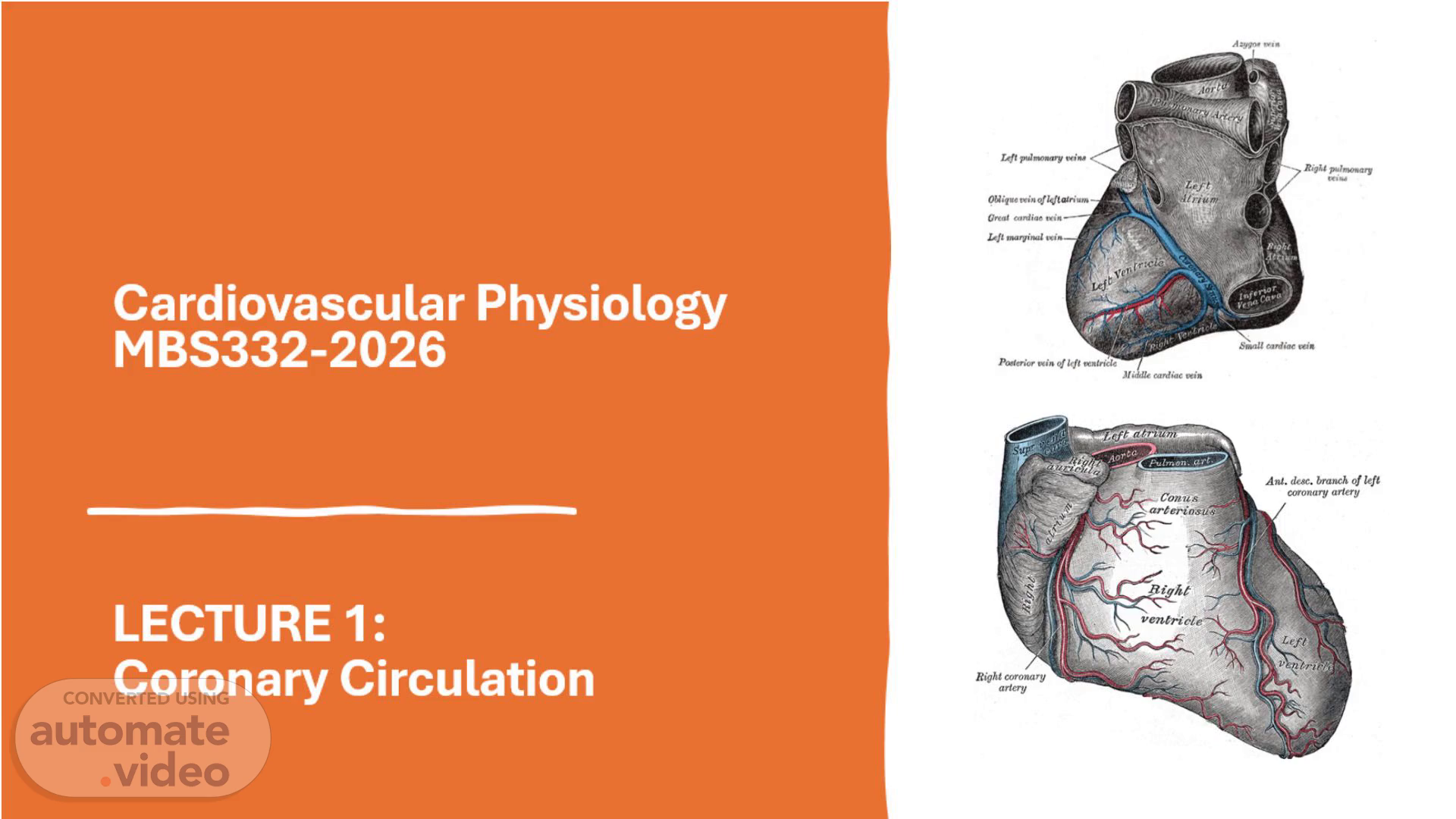
Cardiovascular Physiology MBS332-2026 LECTURE 1: Coronary Circulation.
Scene 2 (15s)
[Audio] Summary: The coronary arteries are the network of blood vessels that branch off the aorta and wrap around the outside of the heart. Their unique function is to deliver freshly oxygenated blood and nutrients to the heart muscle to sustain the heart's O-W-N high demand for oxygen. The two main vessels are the left coronary artery and the right coronary artery, which further divide into smaller branches to supply every part of the myocardium. If these arteries become narrowed or blocked by plaque, it reduces blood flow and can lead to chest pain, heart failure, or a heart attack..
Scene 3 (52s)
[Audio] Learning outcomes – The student must be able to: 1-Give the main coronary vessels and their respective branches, as well as which parts of the heart the vessels supply. 2-Explain in detail coronary dominance (Right against Left) 3-Describe the unique blood flow dynamics (Diastolic perfusion).
Scene 4 (1m 14s)
[Audio] 4-Describe Myocardial Oxygen extraction. 5-Explain what happens when this physiology is interrupted by atherosclerosis or vasospasms. 6-Describe the autoregulation of the coronary arteries (metabolic, myogenic and endothelial). 7-Describe the coronary microcirculation..
Scene 5 (1m 37s)
[Audio] LO 1 The two main coronary arteries arising from the aorta are the Left Coronary Artery (L-C-A--) and the Right Coronary Artery (R-C-A--). They deliver oxygenated blood to specific regions of the heart muscle (myocardium). 1. Left Coronary Artery (L-C-A--) The L-C-A travel a short distance as the Left Main Coronary Artery before splitting into two primary branches. It primarily supplies the left side of the heart. A-Left Anterior Descending (L-A-D--) Artery Areas Supplied: Anterior wall of the left ventricle, anterior two thirds of the interventricular septum, and the apex of the heart. B-Left Circumflex (LCx) Artery Areas Supplied: Lateral wall of the left ventricle, posterior wall of the left ventricle, and the left atrium..
Scene 6 (2m 36s)
[Audio] 2. Right Coronary Artery (R-C-A--) The R-C-A travels along the right coronary sulcus and primarily supplies the right side of the heart and the electrical conduction system. A Right Marginal Artery Areas Supplied: Anterior and lateral walls of the right ventricle. Posterior Descending Artery (P-D-A--) Note: Arises from the R-C-A in 85% of people (right dominant hearts). Areas Supplied: Posterior third of the interventricular septum, inferior wall of both ventricles, and the posteromedial papillary muscle. B-Sinoatrial (S-A---) Nodal Artery (Arises from R-C-A in ~60% of people) Areas Supplied: SA node (the heart's natural pacemaker). C-Atrioventricular (A-V---) Nodal Artery (Arises from RCA in ~80% of people) Areas Supplied: AV node..
Scene 7 (3m 43s)
[Audio] Coronary veins The Greater Cardiac Venous System (Main Surface Veins) Most of the deoxygenated blood from the heart travels through these large, visible surface veins. The Coronary Sinus The coronary sinus is a wide, channel like venous structure located on the back (posterior side) of the heart in the atrioventricular groove. It collects roughly 85% of the heart's total venous blood and empties it directly into the right atrium. It is formed by the confluence of several major surface veins: Great Cardiac Veins: The largest tributary, running along the front of the heart alongside the left anterior descending (L-A-D--) artery. It drains the anterior aspect of both ventricles and the left atrium. Middle Cardiac Veins: Located on the bottom/posterior surface of the heart alongside the posterior descending artery (P-D-A--), draining the back of the ventricles. Small Cardiac Veins: Positioned on the lower right margin of the heart next to the right marginal artery, draining the right atrium and ventricle. Posterior Vein of the Left Ventricle: Runs up the outer left wall to drain the thick left ventricular muscle..
Scene 8 (5m 2s)
[Audio] The Anterior Cardiac Veins These are a distinct group of 3 to 5 small veins on the front of the right ventricle. Unlike the other surface veins, they bypass the coronary sinus entirely and dump blood directly through the front wall of the right atrium. 2. The Smaller Cardiac Venous System (Deep Veins) Thebesian Veins (Venae Cordis Minimae): These are microscopic, hidden veins found deep within the muscular walls of all four heart chambers. Instead of traveling along the surface, they drain blood directly out of the myocardium straight into the nearest internal heart cavity. Because they can empty deoxygenated blood into the left atrium and left ventricle, they contribute to a tiny, normal physiological "shunt" of unoxygenated blood mixing into the systemic arterial loop..
Scene 9 (5m 56s)
[Audio] L-O-2 Coronary artery dominance An anatomical classification that defines which main coronary artery supplies the posterior descending artery (P-D-A--)—the vital vessel running along the back of the heart that feeds the inferior third of the interventricular septum and parts of the left and right ventricles. This layout is categorized into right dominance (70%–85% of the population), left dominance (5%–10%), and co dominance or balanced circulation (5%–20%).
Scene 10 (6m 34s)
[Audio] L-O-3 Cardiac Perfusion Left Ventricle (Primarily Diastole) The Phase: Relaxation (diastole). The Reason: The left ventricle is thick and generates high pressure during contraction (systole). The Mechanism: This strong contraction squeezes the internal blood vessels (coronary arteries) completely shut. Blood can only flow freely when the muscle relaxes. Right Ventricle (Both Systole and Diastole) The Phase: Continuous (both systole and diastole). The Reason: The right ventricle is thin and generates much lower pressure. The Mechanism: The pressure inside the right ventricle during contraction is lower than the pressure in the aorta. Because of this, blood keeps flowing through the right coronary artery during the whole cardiac cycle..
Scene 11 (7m 31s)
[Audio] Key Factors affecting perfusion Heart Rate: Fast heart rates shorten diastole, reducing left ventricle perfusion time. Aorta Pressure: High pressure in the aorta drives blood into the heart muscle. Tissue Depth: The innermost layer of the left ventricle (subendocardium) is the most vulnerable to poor blood flow because it faces the highest squeeze during contraction..
Scene 12 (7m 59s)
[Audio] L-O-4 Myocardial oxygen extraction Is the process by which the heart muscle pulls oxygen out of the blood traveling through the coronary arteries. The heart has the highest resting oxygen extraction rate of any major organ in the body, pulling out 70% to 80% of the oxygen delivered to it even when you are completely at rest..
Scene 13 (8m 26s)
[Audio] L-O-4 The Key Problem: No "Extraction Reserve" Because the heart already extracts nearly all available oxygen at rest, it has almost no extraction reserve. Other Organs: The rest of the body averages an oxygen extraction ratio of only 25% to 30% at rest. If a skeletal muscle works harder, it can simply extract more oxygen from its existing blood supply. The Heart: The heart cannot just "pull more oxygen" when it works harder because it is already running near maximum efficiency..
Scene 14 (9m 1s)
[Audio] How the Heart Compensates Because the extraction percentage cannot go up significantly, the only way the heart can get more oxygen is by increasing coronary blood flow. During heavy exercise or stress, the coronary blood vessels must dilate to physically pump up to 4 to 5 times more blood into the heart muscle. L-O-5 Clinical Significance This unique physiology explains why the heart is highly vulnerable to certain conditions (Interrupted myocardial oxygen extraction): Coronary Artery Disease: If an artery is blocked by plaque, blood flow cannot increase to meet the demand, causing immediate ischemia (angina or a heart attack). Severe Anaemia: A drop in hemoglobin means less oxygen is delivered per milliliter of blood. Because the heart cannot extract a higher percentage to make up for it, ischemia can happen even with normal blood vessels. Tachycardia: A racing heart massively increases oxygen demand while simultaneously shortening the diastolic perfusion time needed to deliver that blood..
Scene 15 (10m 8s)
[Audio] L-O-6 Autoregulation of the coronary arteries (metabolic, myogenic and endothelial) Myogenic Control: Stretch sensitive vascular smooth muscle in the prearterioles constricts in response to high pressure. This maintains a steady blood flow despite changes in systemic blood pressure . Metabolic Regulation: When the heart muscle works harder, cells release vasodilator metabolic byproducts like adenosine, nitric oxide, and hydrogen ions [1, 4]. These compounds signal the arterioles to dilate, rapidly increasing local blood flow . Endothelial Regulation: The inner lining of the microvessels senses frictional forces (shear stress) from blood flow and releases relaxing factors to fine tune the vessel diameter ..
Scene 16 (11m 9s)
[Audio] L-O-7 Coronary microcirculation refers to the vast network of prearterioles, arterioles, and capillaries—all under 500 micrometres in diameter—that regulate blood flow within the heart muscle . While the large epicardial coronary arteries (the ones visible on the surface of the heart) act as conduit vessels, the microcirculation accounts for 90% of coronary vascular resistance and directly controls myocardial tissue perfusion.