Scene 1 (0s)
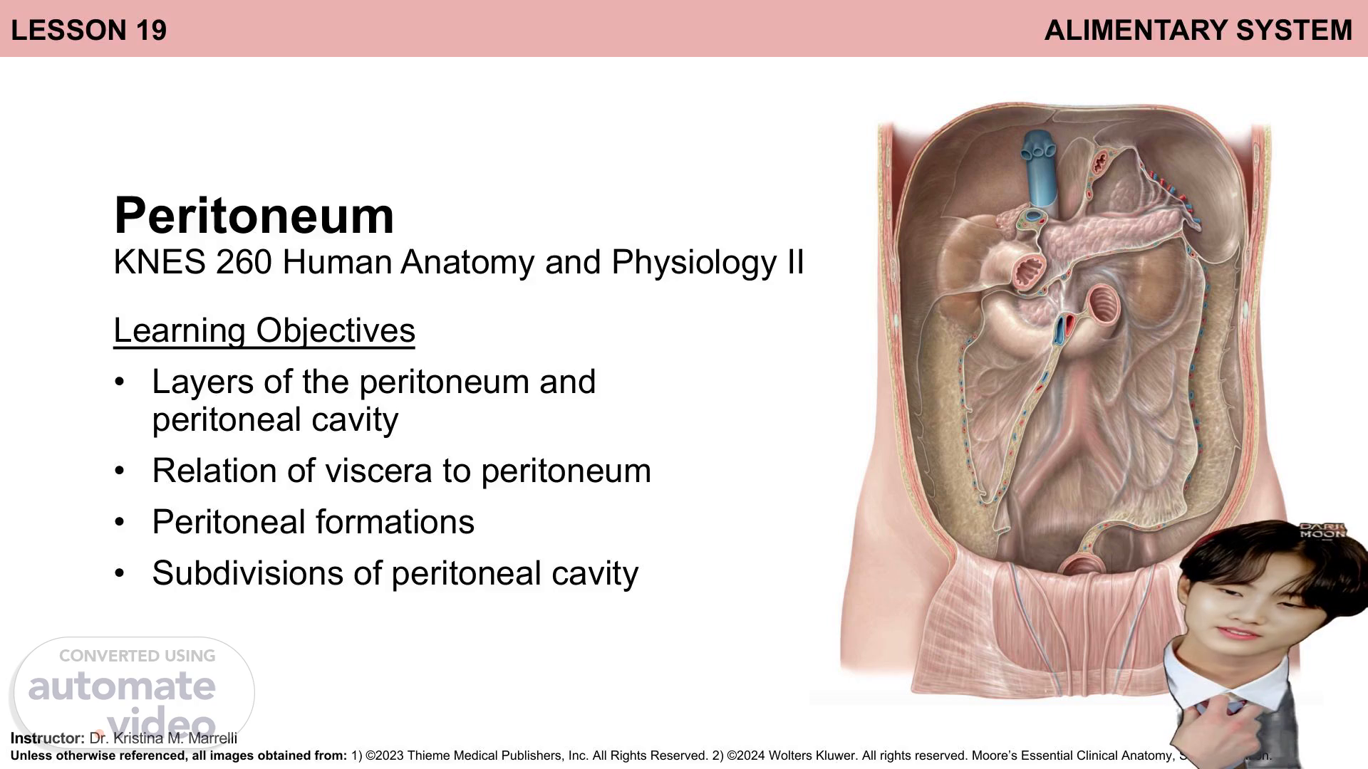
Lesson 19 alimentary SYSTEM Peritoneum KNES 260 Human Anatomy and Physiology 2 Learning Objectives Layers of the peritoneum and peritoneal cavity Relation of viscera to peritoneum Peritoneal formations Subdivisions of peritoneal cavity Instructor: Dr Kristina M Marrelli Unless otherwise referenced, all images obtained from: 1) ©2023 Thieme Medical Publishers, Inc. All Rights Reserved. 2) ©2024 Wolters Kluwer. All rights reserved. Moore’s Essential Clinical Anatomy, Seventh Edition..
Scene 2 (47s)
[Audio] Learning Outcomes By the end of today’s lesson, you will be able to: Describe the formation of the peritoneal cavity and distinguish layers of the peritoneum Differentiate intra-, retro-, and subperitoneal structures based on relationship with peritoneum Identify peritoneal formations: mesentery, omenta, recesses Describe the formation of peritoneal derived structures Locate and describe the locations of subdivisions of the peritoneal cavity.
Scene 3 (1m 17s)
[Audio] Peritoneum Overview Serous (in other words, fluid secreting) membrane lining the abdominopelvic cavity and most viscera contained within cavity Slippery, transparent 2 continuous layers: Parietal peritoneum – lines internal surface of abdominopelvic cavity Visceral peritoneum – invests and lines viscera (for example, stomach, liver, intestines).
Scene 4 (1m 49s)
[Audio] Peritoneum Development Located within abdominopelvic cavity (in other words, extends into pelvis) Peritoneum Peritoneal cavity Lateral view of abdominal and pelvic osteology.
Scene 5 (2m 4s)
[Audio] Peritoneum Parietal and visceral Parietal peritoneum reflects upon itself on the surface of viscera, Intraperitoneal forming the visceral peritoneum structures Retroperitoneal structures Subperitoneal structures Lateral view of abdominal and pelvic osteology Medial view of abdominopelvic cavity; bisection Parietal peritoneum Visceral peritoneum.
Scene 6 (2m 31s)
[Audio] Peritoneum Peritoneal Cavity Potential space between parietal and visceral peritoneum; contains peritoneal fluid that: keeps surfaces moist allows for free movement of viscera (for example, during digestion) contains immune cells to resist infection Peritoneal cavity Parietal peritoneum Medial views of abdominopelvic cavity; bisections Visceral peritoneum.
Scene 7 (2m 58s)
[Audio] Peritoneum Intra-, retro-, sub peritoneal structures Retroperitoneal Subperitoneal Category Intraperitoneal Structures Structures Structures Almost completely External to the parietal External to the parietal covered by visceral peritoneum peritoneum (in other words, outside of the peritoneal cavity) peritoneum (in other words, outside of the peritoneal cavity) Invaginated (in other words, Usually partially covered Usually partially covered protruded) closed sac Anchored by by parietal peritoneum on anterior surface by parietal peritoneum on superior surface Relationship of viscera to peritoneum Posterior To INFERIOR to peritoneal mesentery (doublelayer of peritoneum) peritoneal cavity cavity Mobile structures Esophagus Small intestines: parts of duodenum Distal rectum Urinary bladder Cervix Large intestines: Stomach Spleen Liver Transverse colon Sigmoid Colon Small intestines: jejunum, ileum, part of duodenum ascending colon, descending colon, proximal rectum Example structures Appendix Body of uterus, uterine tubes, ovaries Pancreas (exception: tail) Abdominal aorta Inferior vena cava Kidneys Ureters Medial view of abdominopelvic cavity; bisection.
Scene 8 (4m 38s)
[Audio] Peritoneum Intra-, retro-, sub peritoneal structures Parietal peritoneum Peritoneal cavity Visceral peritoneum Mesentery Intraperitoneal structure Retroperitoneal structure Superior view of abdominopelvic cavity; transverse section Medial view of abdominopelvic cavity; bisection.
Scene 9 (5m 7s)
[Audio] Peritoneum Intra-, retro-, sub peritoneal structures Retroperitoneal structure Parietal peritoneum Peritoneal cavity Superior view of abdominopelvic cavity; transverse section Anterior view; focus on retroperitoneal structures.
Scene 10 (5m 27s)
[Audio] Peritoneal Formations Mesentery Intraperitoneal structure Mesentery Parietal peritoneum Peritoneal cavity Visceral peritoneum Superior view of abdominopelvic cavity; transverse section Double layer of peritoneum (straight up!!!!) Suspends / anchors intraperitoneal structures to body wall Pathway for neurovascular structures to / from viscera.
Scene 11 (5m 54s)
[Audio] Peritoneal Formations Mesentery Intraperitoneal structure Mesentery Superior view of abdominopelvic cavity; transverse section Anterior view; small intestines reflected to highlight mesentery.
Scene 12 (6m 8s)
[Audio] Stop And Think! Peritoneal Formations Mesentery 1. What is the first layer pierced by the B-L-U-E pin? 2. What is the first layer pierced by the R-E-D pin? 3. What is the first layer pierced by the yellow pin? Neurovasculature Anterior view; small intestines cut to highlight root OF The mesentery Anterior views; small intestines reflected to highlight mesentery.
Scene 13 (6m 36s)
[Audio] Peritoneal Formations Greater and lesser omenta Lesser curvature of stomach Greater curvature of stomach Omenta (sing. omentum) Double layered extensions or folds of peritoneum that extend from the stomach and proximal part of duodenum to adjacent organs Liver Lesser omentum Duodenum Lesser omentum Double layered peritoneal fold Lesser curvature of stomach and proximal part of duodenum à liver Comprised of two peritoneal ligaments Transverse colon Greater omentum Greater omentum Four layered (2x double layers) peritoneal fold Descends from greater curvature of stomach and proximal part of duodenum Folds back; attaches to transverse colon Apron like Anterior view.
Scene 14 (7m 24s)
[Audio] Peritoneal Formations Lesser omentum Lesser omentum Double layered peritoneal fold Anterior view Comprised of two continuous peritoneal ligaments Hepatogastric ligament: membranous Medial view of abdominopelvic cavity; bisection Hepatoduodenal ligament: thickened free edge (right side; conducts the portal TRIAD).
Scene 15 (7m 50s)
[Audio] 2 Peritoneal Formations Greater omentum Greater omentum Four layered (2x double layered) peritoneal fold Anterior view of greater omentum in situ; reflected in figure 2 Medial view of abdominopelvic cavity; bisection.
Scene 16 (8m 11s)
[Audio] 2 Peritoneal Formations Greater omentum Liver Stomach ` Body wall Greater omentum T C Peritoneum Parietal peritoneum Visceral peritoneum Anterior view of greater omentum in situ; reflected in figure 2 Lateral view; bisection.
Scene 17 (8m 34s)
[Audio] Subdivisions of the Peritoneal Cavity Greater and lesser peritoneal sacs Superior recess of Liver omental bursa lesser S-A-C (in other words, omental bursa) Stomach ` Body wall GREATER SAC T C Inferior recess of omental bursa Greater sac Main and larger part of peritoneal cavity Peritoneum Lateral view; bisection Parietal peritoneum Visceral peritoneum Lesser sac (OMENTAL bursa) Posterior to stomach and lesser omentum; has superior and inferior peritoneal recesses (in other words, pouches).
Scene 18 (9m 13s)
[Audio] Subdivisions of the Peritoneal Cavity Omental bursa Superior recess of Liver Permits free movement of the stomach omental bursa Communicates with greater sac via the omental ForAMEN omental bursa Stomach ` Body wall Omental foramen T C Inferior recess of omental bursa Anterior view of stomach, lesser omentum, and liver Peritoneum STOP AND THINK! The omental foramen is located posterior to which part of the lesser omentum? Lateral view; bisection Parietal peritoneum Visceral peritoneum.
Scene 19 (9m 56s)
[Audio] Subdivisions of the Peritoneal Cavity Omental bursa Omental foramen Omental bursa Anterior view of stomach, lesser omentum, and liver Anterior view; stomach reflected to highlight location of omental bursa.
Scene 20 (10m 13s)
[Audio] What additional vessels (labelled 1, 2) are present within this section? Are these structures intra-, retro-, subperitoneal? Subdivisions of the Peritoneal Cavity Omental bursa Spleen 1 2 Lesser omentum: Pancreas Stomach Hepatoduodenal ligament Hepatogastric ligament Anterosuperior view; transverse section highlighting access to omental bursa.
Scene 21 (10m 46s)
[Audio] STOP AND THINK Identify structures in the cross section 1 3 4 2 Inferior view Viscera / Vessels 1. 2. 3. 4..
Scene 22 (11m 1s)
[Audio] Summary Peritoneum is a continuous serous membrane lining the abdominopelvic cavity (parietal peritoneum) and surrounding the viscera (visceral peritoneum) The peritoneal cavity is located between the parietal and visceral peritoneum; it contains fluid that permits movement of abdominopelvic viscera and contains immune cells Structures that invaginate the peritoneal cavity are intraperitoneal; structures that are located posterior or inferior, but have one surface covered with parietal peritoneum, are known as retro and subperitoneal, respectively Peritoneum connects organs with adjacent organs or the abdominal wall, forming compartments, recesses, and structures such as: Mesentery: double layered peritoneal derived structure suspending intraperitoneal structures and providing means of passage for neurovascular structures to and from viscera Lesser omentum: double layered peritoneal derived structure extending between the lesser curvature of the stomach and proximal part of the duodenum à liver; the hepatoduodenal and hepatogastric ligaments are continuous peritoneal ligaments of the lesser omentum Greater omentum: apron like, four layered (2x double layer) peritoneal derived structure extending between the greater curvature of the stomach and proximal part of the duodenum à transverse colon; this structure hangs anterior to the small intestines The peritoneal cavity is subdivided into a greater sac (predominant subdivision) and lesser sac (omental bursa) located posterior to the stomach and lesser omentum The lesser sac has a superior peritoneal recess (posterior and inferior to liver and diaphragm) and an inferior peritoneal recess (extending inferiorly between the layers of the greater omentum) Communication between the subdivisions occurs via the omental foramen.
Scene 23 (13m 5s)
[Audio] Identify qAbdominopelvic cavity qStomach qGreater curvature qLesser curvature qLiver qTransverse colon qSmall intestines qDuodenum qJejunum qIleum qParietal peritoneum qVisceral peritoneum qPeritoneal cavity qGreater sac qLesser sac (OMENTAL bursa) qOmental foramen qGreater omentum qLesser omentum qHepatoduodenal ligament qHepatogastric ligament qMesentery qDifferentiate: qIntraperitoneal structures qRetroperitoneal structures qSubperitoneal structures.