Scene 1 (0s)
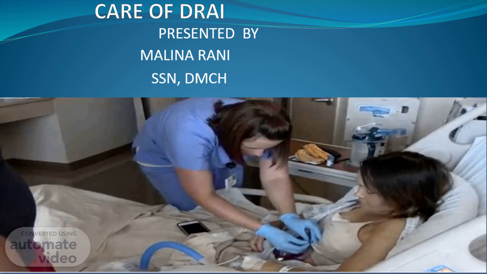
CARE OF DRAIN. MALINA RANI SSN, DMCH. PRESENTED BY.
Scene 2 (8s)
[Audio] The learning objectives of this presentation are to explain the care of drain, discuss the purpose and indication of drain care, explain the classification of drain care, describe the complications and removal of drain care, and explain the nursing care of drain..
Scene 3 (24s)
[Audio] Drain insertion is done to help eliminate dead space, to evacuate existing accumulation of fluid, to remove pus, blood, serous, and bile. This is also done to prevent the potential accumulation of fluid and to prevent infection..
Scene 4 (39s)
[Audio] A drain is a tube used to allow fluid or air to drain freely to the surface from an operation site or wound. This fluid can be blood, serum, pus, urine, feces, bile or lymph..
Scene 5 (52s)
[Audio] Septic wounds should be drained because they have a high risk of developing into abscesses or cellulitis. Aseptic wounds, on the other hand, require careful management to prevent excessive bleeding or fluid accumulation. Wounds with a high likelihood of fluid collection, such as those resulting from operations, need to be monitored closely to prevent complications. Similarly, leaking wounds from anastomosis require prompt attention to prevent further damage. Finally, injuries such as strokes, accidents, or head injuries can lead to significant fluid accumulation and require immediate medical attention..
Scene 6 (1m 29s)
[Audio] A drain system that is connected directly to pressure vessels is called a "pressure" or closed drain system. This type of drain system is formed by tubes into a bag or bottle, such as N G tubes, Foley's catheter, chest, abdominal and orthopedic drains. These types of drains are used when there is a need to collect fluids under pressure, such as in surgical procedures where fluids need to be collected quickly..
Scene 7 (1m 56s)
[Audio] Active drains are closed systems that collect fluid into a reservoir. They apply an artificial pressure to pull fluid or gas from a wound or body cavity. This type of drain is used when there is a need to remove large amounts of fluid quickly. For example, in cases where there is a lot of bleeding or swelling. On the other hand, passive drains have no suction. They rely on pressure differentials, overflow and gravity between body cavities and the exterior to allow fluid to flow out. This type of drain is used when there is a slow and steady flow of fluid needed. For instance, in cases where there is a small amount of fluid leaking from a wound..
Scene 8 (2m 36s)
[Audio] Fluids such as urine, blood, bile, pus, and other bodily secretions are drained from the body through various types of drainage systems. These include NG tubes, Foley's catheters, chest tubes, T-tubes, drains, Penrose drains, pigtail drains, and Redon drains, as well as Jackson-Pratt drains..
Scene 9 (2m 55s)
[Audio] The nasogastric tube, also known as an NG tube, is a flexible tube inserted through the nose and into the stomach. Its primary function is to deliver nutrients, medications, and fluids directly into the stomach, bypassing the mouth and esophagus. This tube can be used for various purposes, including providing nutrition support, administering medication, and managing gastrointestinal issues. Proper placement and use of the NG tube require regular flushing to maintain its functionality and prevent complications..
Scene 10 (3m 26s)
[Audio] A urinary catheter is a hollow, partially flexible tube that collects urine from the bladder and leads to a drain. This type of catheter is typically used when someone is unable to empty their bladder on their own, whether due to retention of urine, incontinence, or loss of bladder control. The catheter serves as a means of collecting and draining urine from the body, allowing individuals to manage their urinary function more effectively..
Scene 11 (3m 50s)
[Audio] A chest tube is a flexible plastic tube, also known as an intercostal catheter, inserted through the chest wall and into the plural space to remove air, fluid, or pus from the thoracic space. Its primary indications include pneumothorax, hemothorax, hemopneumothorax, emphysema, and pleural effusion..
Scene 12 (4m 13s)
[Audio] T-Tube is a type of tube that resembles the letter T, with its top portion inserted within a tubular structure, such as the common bile duct. This device is used for purposes including decompression and cholecystectomy..
Scene 13 (4m 28s)
[Audio] The Penrose drain is a type of drainage device that serves multiple purposes. It not only removes fluid from the wound area, but it can also be used to drain cerebrospinal fluid in patients suffering from hydrocephalus. Additionally, it is commonly employed in various surgical procedures such as plastic surgery, breast surgery, and orthopedic procedures..
Scene 14 (4m 51s)
[Audio] A pigtail catheter is a medical device used to drain urine directly from a kidney. This type of drain allows the fluid to flow freely into the surrounding area around the lungs..
Scene 15 (5m 3s)
[Audio] Redon drains are used to channel secretions, blood, and pus from the wounds or abscess. They function by gravity drainage system..
Scene 16 (5m 15s)
[Audio] A Jackson-Pratt drain, also known as a JP drain, is a type of close suction medical device commonly used as a post-operative drain for collecting bodily fluids from surgical sites..
Scene 17 (5m 30s)
[Audio] Complications can arise during the care of a drain. Inefficient drainage is one such complication, which can occur due to obstruction, poor drain selection, or erosion into hollow organs. Another possible complication is incision dehiscence, which may result from accumulation of fluid, poor placement, or premature removal. These complications need to be recognized and addressed promptly to prevent further harm to the patient..
Scene 18 (5m 56s)
[Audio] Complications related to chest drains can occur due to infection, discomfort, or pain. Infections may arise from ascending bacterial invasion, foreign body reactions, fluid accumulation, or poor post-operative management. On the other hand, discomfort or pain can result from chest tubes with diameters that are too large or stiff tubing. These complications require prompt attention to prevent serious consequences..
Scene 19 (6m 22s)
[Audio] When assessing a drain, it is essential to identify any potential complications. Blockage is one such issue, which can manifest in several ways. For instance, if the temperature of the drain is above 100°F or higher, it may indicate a problem. Additionally, if the drain appears cloudy or emits a foul odor, it could be a sign of blockage. Furthermore, if the drainage suddenly stops despite the drain having previously been functioning properly, it is likely that there is an obstruction present..
Scene 20 (6m 54s)
[Audio] The drainage system inserted is based on only the needs of the patient, and surgeon's preference. The drainage needs to be documented at a minimum of 8 hours and more frequently if the output is high. Notify the on-duty doctor. Drains should be removed once the drainage has stopped or becomes less than about 40ml/day..
Scene 21 (7m 14s)
[Audio] Slide 28 PATIENT EDUCATION. Drains should be removed once the drainage has stopped or becomes less than about 25ml/day Warn the patient that there may be some discomfort when the drain is pulled out. Ensure plan for removal of drain tubing. Explain the patient/parent about removal and possible pain..
Scene 22 (7m 30s)
[Audio] When removing a surgical drain, it is essential to use standard aseptic technique to prevent infection. Clean around the site where the drain is located, making sure to remove any sutures that may be present. Pinch the edges of the skin together and gently rotate the tubing from side to side to loosen the drain. Then, remove the drain smoothly but quickly, taking care not to pull on the surrounding tissue. Once the drain is removed, place a dry dressing over the site to keep it clean and protect it from further irritation. Finally, document the patient's progress notes to track their recovery..
Scene 23 (8m 6s)
[Audio] When assessing the drain insertion site, it is essential to look for any signs of leakage, redness, or oozing. This is crucial to prevent complications and ensure proper healing. To secure the drain, sutures or tapes may be used. It is vital to document the site condition and inform the on-duty doctors about any issues found. Additionally, we need to ensure that the drain is positioned below the insertion site and free from any kinks or knots. This will help maintain its effectiveness and reduce the risk of complications..
Scene 24 (8m 36s)
[Audio] The amount and type of fluid in the drain bottle must be noted and documented. If the patient has a fever, redness, tenderness, or increased oozing at the drain site, monitor them closely for signs of sepsis. An infection may be present, prompting notification of the responsible doctors and potentially requiring blood cultures to be taken..
Scene 25 (8m 57s)
[Audio] The drain patency and insertion site should be observed regularly. This includes checking whether the drain is functioning properly and whether there are any signs of infection or irritation at the insertion site. If suction is required, it should be ensured that it is maintained correctly. Additionally, drainage needs to be monitored closely, documenting the amount and type of fluid being drained at least every four hours, and more frequently if the output is high. Furthermore, the insertion site should be cleaned daily with sterile saline solution to prevent infection..
Scene 26 (9m 32s)
[Audio] When emptying the collection bulb on the drain three times daily, it is essential to do so carefully to avoid any complications. This process helps to prevent the buildup of fluid and reduces the risk of infection. Moreover, it is crucial to monitor the drain closely, as a blocked drain tube can lead to severe consequences such as the formation of a hematoma, increased pain, and even infection. Therefore, it is vital to remove the drain under proper observation to minimize these risks..
Scene 27 (10m 2s)
[Audio] The patient or parent should be educated to ensure the drain is positioned below the site of insertion, but not pulling on the patient. There is a risk of dislodgement, which requires increased care when moving. Moving while the drain is in place may cause some pain, but this can be minimized with regular analgesia..
Scene 28 (10m 21s)
[Audio] The references provided emphasize the significance of educating patients or parents about the proper care of drains. This involves ensuring the drain is positioned correctly, being mindful of the risk of dislodgement, and taking measures to minimize discomfort while the drain is in place. By offering clear instructions and guidance, healthcare professionals can enable individuals to take an active role in their own care and decrease the probability of complications..
Scene 29 (10m 47s)
[Audio] The fluids to be drained from the body are blood, serum, pus, urine, feces, bile, or lymph..
Scene 30 (11m 11s)
[Audio] The fluids to be drained from body may include blood, serum, pus, urine, feces, bile, or lymph. The drainage system inserted is based on only the needs of patient, and surgeon preference. Drainage needs to be documented at a minimum 8 hourly and more frequently if output is high and notify on duty doctors. Drains should be removed once the drainage has stopped or becomes less than about 40ml/day. Passive drains are maintained under suction. To assess a drain which is inserted in body, one should assess the drain insertion site for signs of leakage, redness or sign of ooze, document site condition and notify on duty doctors. One should also assess if drain is secured with suture or tape, document it, assess potency of drain, ensure drain is located below the insertion site and free from kinks or knots. Note and document amount and type of fluid in drain bottle. Monitor patient for signs of sepsis if the patient is febrile, has redness, tenderness, or increased ooze at the drain site, this could be a sign of infection, then inform the responsible doctors and blood cultures may need to be obtained..
Scene 31 (12m 20s)
THANKS.
