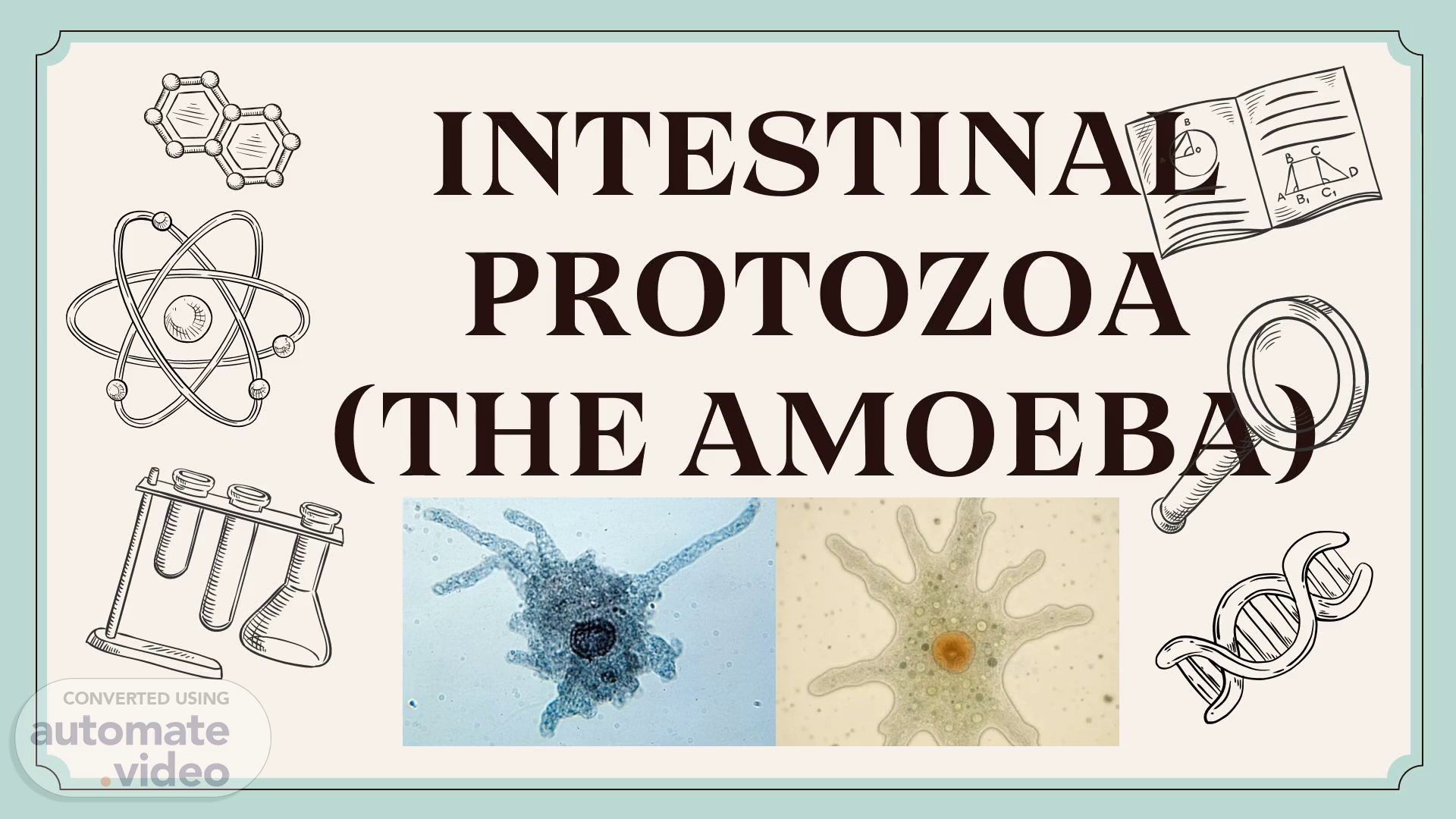
Scene 1 (0s)
[Audio] Entamoeba histolytica is a parasitic protozoan that causes disease in humans. The parasite has two main types of forms: trophozoites and cysts. Trophozoites are the active form of the parasite, responsible for invading host cells and causing damage to tissues. Cysts are the dormant form of the parasite, which can survive outside of a host for extended periods of time. Both forms have distinct morphological features that allow them to be identified. The pathogenicity of E. histolytica is primarily due to its ability to invade host cells and cause tissue damage. This invasion leads to inflammation and necrosis of infected tissues, resulting in symptoms such as diarrhea, abdominal pain, and weight loss. The parasite also produces enzymes that help it to break down host tissues. E. histolytica is typically found in the large intestine, where it feeds on nutrients from food. However, it can also be found in other parts of the body, including the liver and spleen. The parasite can infect both healthy individuals and those with compromised immune systems. The classification of E. histolytica is based on its morphology and genetic characteristics. It belongs to the phylum Apicomplexa, class Stramenopodia, order Valentimorpha, family Entamoebidae, genus Entamoeba, and species E. histolytica. Its genetic makeup is unique and allows it to adapt to different environments. The distribution of E. histolytica varies depending on the region and climate. It is most commonly found in tropical and subtropical regions, particularly in areas with poor sanitation and hygiene. In these regions, the parasite can spread quickly through contaminated water and food sources. Morphologically, E. histolytica can be distinguished by its elongated shape, pointed ends, and distinctive nuclei. The trophozoite form has a characteristic "V"-shaped nucleus, while the cyst form has a rounded nucleus. These morphological differences allow for accurate identification of the parasite. Identification of E. histolytica requires specialized equipment and techniques. Microscopic examination of stool samples is often used to detect the presence of the parasite. Other methods include culturing the parasite in a laboratory setting and using molecular biology techniques to analyze its genetic material..
Scene 2 (2m 38s)
[Audio] ## Step 1: Identify the main topic The main topic is about intestinal protozoa, specifically the classification of amoebas. ## Step 2: Determine the classification of amoebas There are nine different types of amoebas classified as follows: Entamoeba histolytica, Entamoeba coli, Entamoeba dispar, Entamoeba hartmanii, Entamoeba moshkovskii, Entamoeba poleckii, Entamoeba gingivalis, Endolimax nana, and Iodomaoeba butschiii. ## Step 3: Explain the characteristics of each type Each of these classifications has its own unique characteristics that play a crucial role in the human digestive system. ## Step 4: Discuss the importance of intestinal protozoa Intestinal protozoa, including the amoebas mentioned above, are essential for maintaining the balance of the human digestive system. The final answer is:.
Scene 3 (3m 41s)
[Audio] The pathogenic intestinal amoeba are single-celled organisms that live in various environments, including water and soil. Most amoebas are harmless, but some can cause serious health issues when they infect the human intestines. These amoebas are commonly found in tropical and subtropical regions and can be transmitted through contaminated food or water. They enter the body through the mouth and then travel to the intestines, causing a range of symptoms. Diarrhea, abdominal pain, cramping, and bloody stools are among the most common symptoms of a pathogenic intestinal amoeba infection. In severe cases, the infection can lead to serious complications like liver abscesses or even death. Not everyone who comes into contact with these amoebas will become ill, but people with weakened immune systems, such as young children, older adults, and individuals with underlying health conditions, are at a higher risk of developing a serious infection. To prevent the spread of pathogenic intestinal amoeba, it is essential to practice good hygiene, such as washing your hands before and after handling food, and drinking clean and safe water. Avoiding consumption of food or drinks from questionable sources and cooking meat thoroughly to kill any potential amoebas is also crucial. The prevention of pathogenic intestinal amoeba infections requires awareness and caution..
Scene 4 (5m 14s)
[Audio] The parasite Entamoeba histolytica causes intestinal infections by invading the mucosa of the small intestine. The invasion leads to inflammation and damage to the intestinal lining. The symptoms of this infection are similar to those caused by other gastrointestinal diseases such as gastritis and gastroenteritis. However, the presence of blood in the stool is a distinctive feature of Entamoeba histolytica infection. The disease can be spread through contaminated food and water, fecal-oral transmission, and contact with infected individuals..
Scene 5 (5m 52s)
[Audio] The amoeboid cell body is characterized by its ability to change shape in response to environmental stimuli. This characteristic allows it to move towards food sources and avoid predators. The movement of the amoeboid cell body is achieved through a process called pseudopodia formation. Pseudopodia are extensions of the cell membrane that protrude from the cell body and allow the cell to engulf food particles. The amoeboid cell body can also form a structure called a ventral disc, which helps to anchor the cell to the substrate. The amoeboid cell body has several characteristics that distinguish it from other types of cells. One of these characteristics is its ability to undergo a process called endocytosis. Endocytosis is the process by which a cell engulfs external substances, such as food particles or pathogens. In the case of the amoeboid cell body, endocytosis plays a critical role in the digestion of food particles. Another characteristic of the amoeboid cell body is its ability to regulate its internal pH levels. The amoeboid cell body can maintain a stable internal pH level despite changes in the external environment. This ability is essential for the survival of the cell, as an unstable internal pH level can lead to cellular damage. The amoeboid cell body also has a unique way of moving around. Unlike other cells, the amoeboid cell body does not use traditional muscle contractions to move. Instead, it uses a process called "amoeboid locomotion", where it slowly moves forward by extending and retracting its pseudopodia. This type of movement allows the cell to navigate through complex environments and avoid obstacles. In addition to its unique movement mechanism, the amoeboid cell body also has a specialized structure called a "cilia". Cilia are hair-like structures that protrude from the surface of the cell and help to capture food particles. The cilia of the amoeboid cell body are highly sensitive and can detect even slight changes in the surrounding environment. Overall, the amoeboid cell body is a remarkable example of evolutionary adaptation, with its unique characteristics allowing it to thrive in a wide range of environments. Its ability to regulate its internal pH levels, engage in endocytosis, and utilize amoeboid locomotion makes it an ideal candidate for studying cellular biology..
Scene 6 (8m 27s)
[Audio] The trophozoite stage of Entamoeba histolytica is characterized by its size, which can range from 12 to 60 micrometers in diameter. The trophozoite is an anaerobic parasite, meaning it thrives in environments without oxygen. This stage is responsible for causing invasive infections in humans, as well as being the feeding and replication form found in the feces of patients with active disease. The trophozoite has a unique cytoplasm, which is divided into clear ectoplasm and granular endoplasm. The surface of the trophozoite is covered in phagocytic stomata, which are used for engulfing food. The trophozoite's diet consists of freshly ingested erythrocytes, or red blood cells, which appear as pale greenish, refractile bodies within the trophozoite. The trophozoite has the ability to form pseudopodia, which are cytoplasmic protrusions used for movement and engulfing food. These protrusions are characteristically hyaline when first formed. The trophozoite lacks certain organelles found in most cells, such as mitochondria, endoplasmic reticulum, and the golgi apparatus. This is due to its unique metabolism and adaptation to its environment. The trophozoite is an essential part of the life cycle of Entamoeba histolytica..
Scene 7 (9m 53s)
[Audio] The amoeba Entamoeba histolytica was first identified in 1878 by German scientist Rudolf Leuckart. He described it as a parasite that causes disease in humans. Since then, numerous studies have been conducted to better understand its behavior and life cycle. One study published in 2011 found that Entamoeba histolytica can infect both humans and animals. Another study published in 2013 found that the amoeba can survive outside of a host for extended periods of time. These findings suggest that Entamoeba histolytica may not be limited to being a human pathogen, but could also potentially affect other animals..
Scene 8 (10m 18s)
[Audio] The nucleus of the Entamoeba histolytica is located at the center of the cell. The nucleus is surrounded by a clear cytoplasm that contains various organelles necessary for cellular functions. The nucleus itself is made up of chromatin, which is composed of DNA and proteins. The chromatin is organized into distinct regions called nucleoli, where transcription occurs. The nucleoli are responsible for producing messenger RNA, which carries genetic information from the nucleus to the rest of the cell. The nucleus also contains other essential components like mitochondria, which generate energy for the cell. The presence of mitochondria indicates that the cell has the ability to metabolize nutrients and produce energy. The nucleus plays a critical role in the survival and reproduction of the Entamoeba histolytica. Without it, the cell would not be able to function properly. The nucleus is essential for the cell's ability to carry out its biological processes, including growth, development, and reproduction. The nucleus is also involved in regulating the expression of genes, which determines the cell's phenotype and behavior. In summary, the nucleus is a vital component of the Entamoeba histolytica, and its proper functioning is essential for the cell's overall health and well-being..
Scene 9 (11m 48s)
[Audio] The life cycle of Entamoeba histolytica includes both free-living and parasitic phases. The parasite exists in three main forms: the trophozoite, the cyst, and the excystate. The trophozoite is the active feeding stage, while the cyst is the dormant stage that allows the parasite to survive outside of its host. The excystate is the intermediate stage that occurs during the transformation from the cyst to the trophozoite. During the excystation process, the cyst wall breaks down and releases the trophozoite inside. The trophozoite then feeds on nutrients and grows until it reaches maturity. At this point, it transforms back into a cyst through a process called excystation. The entire life cycle of Entamoeba histolytica takes approximately 2 weeks to complete. The parasite can infect various tissues including the liver, spleen, and small intestine. The symptoms of infection may include abdominal pain, diarrhea, weight loss, and fatigue. Treatment typically involves antiparasitic medications such as metronidazole or albendazole..
Scene 10 (13m 0s)
[Audio] The nucleus of the amoeba is crucial for its survival, and its structure is similar to that of the trophozoite when stained. However, the karyosome, or the central portion of the nucleus, is eccentric, meaning it is not located at the center. This unique characteristic helps distinguish the amoeba from other organisms. The cytoplasm contains rod-shaped structures called chromatoidal bars, which surround a glycogen vacuole. These bars do not stain with iodine, but turn a light yellowish-green to yellow-brown color when stained with hematoxylin. They appear as bluish-black when stained with trichrome stain, and as a bright red color. The combination of these characteristics allows us to identify the Entamoeba histolytica species..
Scene 11 (13m 52s)
[Audio] The company has been operating for over 50 years, with a history that spans multiple generations. The company's founder was a visionary who had a clear vision for his business. He believed that innovation and progress were essential to success. He invested heavily in research and development, which enabled him to stay ahead of the competition. His leadership style was characterized by a strong sense of integrity and a commitment to excellence. He fostered an environment where employees could grow and develop their skills. He also encouraged innovation and creativity within the organization. The company's current CEO is a direct descendant of the founder. She has continued the legacy of her ancestor by prioritizing innovation and progress. She has implemented various initiatives to drive growth and improve efficiency. Her leadership style is built on a foundation of trust, respect, and open communication. She has created a culture of collaboration and teamwork, which has led to significant improvements in employee satisfaction and productivity. The company continues to thrive under her leadership, driven by its core values of innovation, progress, and excellence..
Scene 12 (15m 9s)
[Audio] The intestinal protozoan Entamoeba histolytica causes infections in humans. The parasite's presence must be determined in order to accurately diagnose and treat the disease. Microscopy is one method of diagnosis, allowing identification of the parasite's trophozoite stage under a microscope. Stool culture is another method, examining the stool for the parasite's presence. Antigen detection using ELISA can also identify the parasite. This method detects specific proteins produced by the parasite. Antibody detection can be used through various methods, including indirect immunofluorescence assay, ELISA, complement fixation test, and counter immunoelectrophoresis. Histopathology involves examining tissue samples to detect the parasite's presence. Biopsy is used to take tissue samples that are then examined under a microscope. Molecular diagnosis has become more accessible with advancements in technology, particularly through PCR. This method detects the parasite's genetic material and is highly sensitive and specific. Ultrasonography can also be used to diagnose amoebic liver abscess, a complication of Entamoeba histolytica infection. This imaging technique shows the presence of abscesses in the liver, often caused by the parasite. Various diagnostic methods are available, including microscopy, stool culture, antigen and antibody detection, histopathology, molecular diagnosis, and ultrasonography. Accurate diagnosis is essential to provide appropriate treatment and prevent complications..
Scene 13 (16m 50s)
[Audio] The Entamoeba histolytica parasite causes disease in humans through the ingestion of contaminated food or water. The parasite's life cycle includes both free-living and parasitic stages. The free-living stage occurs when the parasite is present in the environment, where it multiplies and produces cysts. The parasitic stage occurs when the parasite infects a human host, where it invades tissues and causes damage. The parasite's life cycle also involves the production of enzymes that help break down tissue. The transmission of E. histolytica from one person to another typically occurs through the fecal-oral route. Fecal matter containing the parasite's cysts is ingested, usually through contaminated food or water. The parasite's cysts can survive for extended periods outside of the human body, allowing for the potential for long-term transmission. To prevent the spread of E. histolytica, it is essential to identify and treat asymptomatic carriers who may be shedding cysts in their stool. Asymptomatic individuals who have passed cysts in their stool should be treated with medications such as metronidazole and tinidazole. Additionally, proper hygiene practices, such as washing hands regularly, can help reduce the risk of transmission..
Scene 14 (18m 15s)
[Audio] The intestinal protozoa, specifically the Amoeba, are discussed in slide 14 of the presentation. The non-pathogenic intestinal amoebas are a type of single-celled organism found in the human and animal intestines. They are commonly found in soil, water, and other environmental sources. Non-pathogenic intestinal amoebas do not cause disease or harm to humans. Instead, they play an important role in the ecosystem by breaking down organic matter and aiding in nutrient cycling. Some amoebas can cause illness, but these particular types are harmless. However, being aware of the presence of amoebas in our environment is crucial. They can be easily transmitted through contaminated food or water, causing symptoms like diarrhea, cramps, and abdominal pain. To prevent the spread of these organisms, proper hygiene and sanitation practices must be followed. In rare cases, non-pathogenic amoebas can become pathogenic, posing a risk to individuals with weakened immune systems or underlying health conditions. If symptoms persist or worsen, seeking medical attention is essential. Non-pathogenic intestinal amoebas are a common and harmless presence in our environment. Taking necessary precautions is vital to prevent illness..
Scene 15 (19m 44s)
[Audio] The Entamoeba coli parasite is found in the human body and in various environments such as freshwater and marine waters, soil and sediments, as well as manure and vegetation. It has three main forms: the trophozoite, precyst, and cyst. The trophozoite form is the active form of the parasite and is capable of causing infection and harm to the host. The precyst form is a transitional stage between the trophozoite and the cyst, which is the dormant and highly resistant form of the parasite. The mode of transmission for Entamoeba coli is through the fecal-oral route. This means that the parasite is transmitted through the ingestion of contaminated food or water that has come into contact with fecal matter containing the parasite. Entamoeba coli can be found in the lower intestines, specifically the colon, but it can also be found in other locations in the outside environment. It is essential to be aware of the different forms of this parasite in order to prevent and treat any potential infections..
Scene 16 (20m 52s)
[Audio] The amoebas found in the human gut are primarily parasitic organisms that feed on bacteria and other microorganisms. They have a unique cell structure that allows them to survive in the harsh environment of the gut. The trophozoite stage is the active, feeding stage of the amoeba, characterized by the presence of a nucleus with irregular clumps of peripheral chromatin. The karyosome, or the central part of the nucleus, is large and irregularly shaped, often located in an eccentric position. In contrast, the cyst stage is the dormant and infective form of the amoeba, marked by its distinctive nuclear structure. The most notable feature of the cyst stage is the presence of splinter-shaped or irregular chromatoids. These characteristics enable us to distinguish between different types of amoebas using staining techniques. Proper identification of the amoeba is essential for accurate diagnosis and effective treatment of infections caused by these parasites..
Scene 17 (21m 56s)
[Audio] The habitat for Intestinal Protozoa, particularly the Amoeba, is the large intestine or colon. This is where they thrive and reproduce. Intestinal Protozoa, including Amoeba, are transmitted through the formation of cysts. These cysts can form in the organism's life cycle and can be ingested through contaminated food and water. One particular Amoeba species, E. moshkovskii, thrives in river and sewage environments. This species is morphologically indistinguishable from another species, E. histolytica, except for the presence of ingested red blood cells in the latter. Entamoeba dispar and Entamoeba moshkovskii are found in the trophozoite form. This form is the active and feeding stage of the organism's life cycle. In summary, the intestinal habitat and modes of transmission for Intestinal Protozoa, specifically the Amoeba, are vital aspects to consider when studying and preventing the spread of these organisms. With advancements in technology and methods of analysis, we can continue to learn more about these organisms and take necessary measures to keep them at bay..
Scene 18 (23m 12s)
[Audio] The trophozoite stage of Entamoeba histolytica ranges in size from 3-12 micrometers, while the cyst stage measures between 4-10 micrometers. Its habitat is the lumen of the large intestine, specifically the colon. This means that it resides in our digestive system. The mode of transmission is primarily through the fecal-oral route, where contaminated food or water is ingested. Proper hygiene and sanitation practices are essential to prevent the spread of this parasite. Laboratory diagnosis involves two main methods: direct fecal smear and permanent staining with iron hematoxylin. A direct fecal smear involves examining a sample of feces under a microscope, allowing for an accurate observation of the amoeba. Permanent staining provides a more detailed and accurate observation of the amoeba. Another variant of Entamoeba, Entamoeba hartmanni, exists only in its cyst form and should be considered when diagnosing and treating this parasite. The differences between E. histolytica and E. hartmanni must be taken into account for proper diagnosis and treatment..
Scene 19 (24m 25s)
[Audio] The amoebas that infect humans are typically found in the oral cavity. They are usually harmless but can cause a variety of symptoms if they multiply excessively. The most common species of amoeba that infects humans is Entamoeba histolytica. However, other species like Entamoeba gingivalis can also infect humans. Entamoeba gingivalis is commonly found in the oral cavity and can cause gum disease and other oral health issues. It is difficult to diagnose because it does not produce any visible signs or symptoms until it has multiplied and caused damage. Diagnosis is often made based on the results of a stool test or a biopsy of the affected area. Once diagnosed, treatment involves antibiotics or antiprotozoal medications. Antibiotics may not be effective against all types of amoebiasis, so alternative treatments such as metronidazole may be necessary..
Scene 20 (25m 25s)
[Audio] The Amoeba is a type of Intestinal Protozoa that can be found in the large intestine, also known as the colon. It is transmitted through the fecal-oral route, which means it is ingested through contaminated food or water. To diagnose the presence of Intestinal Protozoa, there are two laboratory methods that can be used: the Concentrated Wet Mount and Permanent Staining. The Concentrated Wet Mount involves observing a small sample of fecal matter under a microscope. Permanent Staining involves preparing a slide with a sample of fecal matter and staining it with certain dyes to make the organisms more visible. The Amoeba has a distinctive nucleus called a karyosome, which is eccentric and irregular, with several achromatic strands extending to the nuclear membrane. The cytoplasm of the Amoeba does not contain a chromatoidal body or glycogen vacuole. The Amoeba has two forms: the trophozoite, which is about 8-10 micrometers in size and has a sluggish motility, and the cyst, which is smaller at 6-8 micrometers and can have 1-4 nuclei. Endolimax Nana is another type of Intestinal Protozoa that has similar characteristics to the Amoeba, but it does not have a cyst form and is only found in the trophozoite form within the human body..
Scene 21 (26m 47s)
[Audio] Iodamoeba butschlii is a type of protozoan parasite that infects the human gut. It is typically found in the large intestine and cecum. The parasite is usually transmitted through the ingestion of its cyst form, via the fecal-oral route. There are two main methods used to diagnose Iodamoeba butschlii in a laboratory setting. One method is through a concentrated wet mount, where a sample of stool is examined under a microscope. Another method is through permanent staining, which provides a more detailed view of the parasite. When viewed under a microscope, a trophozoite of Iodamoeba butschlii measures approximately 12-15 micrometers in size. Its cytoplasm lacks differentiation between ectoplasm and endoplasm, and appears more vacuolated. A cyst of Iodamoeba butschlii is slightly larger, measuring between 10-20 micrometers in size. It has a characteristic "bull's eye" or "basket" appearance due to its central karyosome surrounded by refractile chromatin granules. On a permanent smear, the cyst may appear to have a halo around the karyosome. The cytoplasm of the cyst also contains a large mass of glycogen or an iodophilic body. The parasite does not possess a chromatid body. This information provides a comprehensive understanding of Iodamoeba butschlii..
Scene 23 (28m 21s)
[Audio] The character of "Don't know." was created by the artist and writer, John R. Miller. He first appeared in the "Don't Know" comic strip which ran from 1950 to 1962. The character's popularity grew rapidly as he became a regular feature in various newspapers and magazines. His popularity peaked around 1955 when he was featured on the cover of Time magazine. The character's popularity declined gradually over time due to his lack of development and the increasing competition from other popular comics. By the end of the 1960s, the character had largely disappeared from circulation. The character's design was influenced by the classic cartoon character, Felix the Cat. The character's personality was based on a mix of ignorance and confusion, with a tendency to ask questions that are easily answered by others. His catchphrase was "I don't know", which became a well-known phrase among readers. The character's popularity was also fueled by his ability to elicit reactions from readers, often asking questions that were intentionally vague or ambiguous. This allowed him to become a sort of "everyman" figure, relatable to many people. The character's creator, John R. Miller, was an American cartoonist who worked for several major newspapers and magazines. He was known for his work on various comic strips and cartoons, but "Don't Know" remains one of his most notable creations. Miller's style was characterized by simple yet effective illustrations, which helped to make the character appealing to a wide range of audiences. His use of humor and wit in the character's dialogue added to its appeal, making it a beloved figure among readers. The character's decline in popularity can be attributed to several factors. Firstly, the character's lack of development and evolution over time meant that he failed to keep up with changing reader tastes. Secondly, the increasing competition from other popular comics and cartoons led to a decline in the character's visibility. Finally, the character's inability to adapt to new technologies and media formats made it difficult for him to remain relevant in the modern era. Despite these challenges, the character remains a nostalgic reminder of the past, evoking memories of childhood and simpler times. His enduring popularity is a testament to the power of nostalgia and the human desire to revisit fond memories. The character's legacy continues to inspire new generations of artists and writers, ensuring that his memory lives on in the hearts of fans everywhere..
Scene 24 (31m 6s)
[Audio] The Amoeba is a type of protozoan parasite that causes disease in humans. It is a single-celled organism that lives in the human intestine. The Amoeba is known for its ability to cause severe diarrhea and dysentery in humans. In fact, it is one of the most common causes of intestinal parasites in humans. The Amoeba's ability to infect humans is due to its unique characteristics, which allow it to survive outside of its host. The Amoeba's survival mechanism involves using enzymes to break down proteins and other nutrients. This process allows the Amoeba to obtain essential nutrients from the human body. The Amoeba's infection mechanism involves the use of a specialized structure called a pseudopod. The pseudopod is used to attach itself to the intestinal wall and begin the infection process. Once attached, the Amoeba uses its enzymes to break down the intestinal lining and create an environment conducive to its growth. The Amoeba's growth and reproduction involve complex processes that require specific conditions. The Amoeba's life cycle includes stages of active growth, dormancy, and death. The Amoeba's ability to adapt to different environments makes it a formidable opponent in the fight against intestinal parasites. The Amoeba's unique characteristics make it a significant threat to public health. The Amoeba's impact on human health is evident in the severity of symptoms caused by its infection. The Amoeba's infection can lead to severe diarrhea, dysentery, and even death in extreme cases. The Amoeba's ability to evade detection and treatment makes it a challenging foe for healthcare professionals. The Amoeba's role in spreading diseases is often underestimated, but it plays a significant part in the transmission of intestinal parasites. The Amoeba's influence on human health is undeniable, and its eradication is crucial for maintaining public health..