Structure of the Heart
Scene 1 (0s)
Structure of the Heart. The human heart is a four-chambered muscular organ , shaped and sized roughly like a man's closed fist with two-thirds of the mass to the left of midline. The heart is enclosed in a pericardial sac that is lined with the parietal layers of a serous membrane . The visceral layer of the serous membrane forms the epicardium ..
Scene 2 (19s)
Layers of the Heart Wall. Three layers of tissue form the heart wall. The outer layer of the heart wall is the epicardium, the middle layer is the myocardium , and the inner layer is the endocardium ..
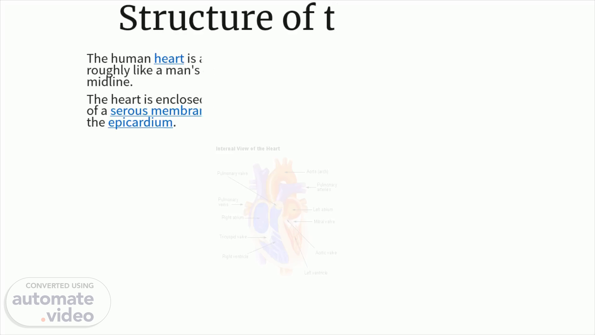
Scene 3 (55s)
Valves of the Heart. Pumps need a set of valves to keep the fluid flowing in one direction and the heart is no exception. The heart has two types of valves that keep the blood flowing in the correct direction. The valves between the atria and ventricles are called atrioventricular valves (also called cuspid valves), while those at the bases of the large vessels leaving the ventricles are called semilunar valves . The right atrioventricular valve is the tricuspid valve. The left atrioventricular valve is the bicuspid, or mitral, valve. The valve between the right ventricle and pulmonary trunk is the pulmonary semilunar valve. The valve between the left ventricle and the aorta is the aortic semilunar valve. When the ventricles contract, atrioventricular valves close to prevent blood from flowing back into the atria. When the ventricles relax, semilunar valves close to prevent blood from flowing back into the ventricles..
Scene 4 (1m 33s)
Pathway of Blood through the Heart. While it is convenient to describe the flow of blood through the right side of the heart and then through the left side, it is important to realize that both atria and ventricles contract at the same time. The heart works as two pumps, one on the right and one on the left, working simultaneously . Blood flows from the right atrium to the right ventricle, and then is pumped to the lungs to receive oxygen . From the lungs, the blood flows to the left atrium, then to the left ventricle. From there it is pumped to the systemic circulation ..
Scene 5 (2m 2s)
Blood Supply to the Myocardium. The myocardium of the heart wall is a working muscle that needs a continuous supply of oxygen and nutrients to function efficiently. For this reason, cardiac muscle has an extensive network of blood vessels to bring oxygen to the contracting cells and to remove waste products. The right and left coronary arteries, branches of the ascending aorta, supply blood to the walls of the myocardium. After blood passes through the capillaries in the myocardium, it enters a system of cardiac (coronary) veins. Most of the cardiac veins drain into the coronary sinus, which opens into the right atrium..
Scene 6 (2m 29s)
Physiology of the Heart. The conduction system includes several components. The first part of the conduction system is the sinoatrial node . Without any neural stimulation , the sinoatrial node rhythmically initiates impulses 70 to 80 times per minute. Because it establishes the basic rhythm of the heartbeat, it is called the pacemaker of the heart . Other parts of the conduction system include the atrioventricular node , atrioventricular bundle , bundle branches, and conduction myofibers . All of these components coordinate the contraction and relaxation of the heart chambers..
Scene 7 (2m 54s)
Cardiac Cycle. The cardiac cycle refers to the alternating contraction and relaxation of the myocardium in the walls of the heart chambers, coordinated by the conduction system, during one heartbeat. Systole is the contraction phase of the cardiac cycle, and diastole is the relaxation phase. At a normal heart rate , one cardiac cycle lasts for 0.8 second..
Scene 8 (3m 19s)
Cardiac Cycle.
Scene 9 (3m 25s)
Heart Rate. The sinoatrial node, acting alone, produces a constant rhythmic heart rate. Regulating factors are reliant on the atrioventricular node to increase or decrease the heart rate to adjust cardiac output to meet the changing needs of the body. Most changes in the heart rate are mediated through the cardiac center in the medulla oblongata of the brain . The center has both sympathetic and parasympathetic components that adjust the heart rate to meet the changing needs of the body. Peripheral factors such as emotions, ion concentrations, and body temperature may affect heart rate. These are usually mediated through the cardiac center..