Development of Heart
Scene 1 (0s)
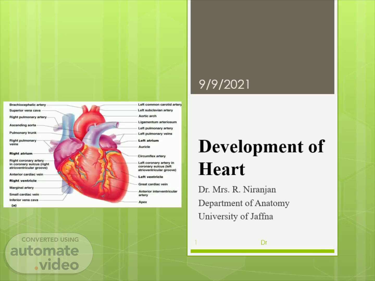
Development of Heart. Dr. Mrs. R. Niranjan Department of Anatomy University of Jaffna.
Scene 2 (12s)
Object i ves. To understand the Establishment of the cardiogenic field Development of primitive heart tube Division and position of the primitive heart Formation of the cardiac loop and its clinical correlates Formation of the coverings of heart Development of the sinus venosus.
Scene 3 (49s)
Development of the cardiovascular system. Angioblasts arise from: Mesoderm Mesenchyme Angioblast cells g ive rise to blood and blood vessels Heart begins to function by end of the 3 rd week - in order to meet the nutrient needs of rapidly growing embryo.
Scene 4 (2m 15s)
ESTABLISHMENT OF THE. 4. CARDIOGENIC FIELD. 9/9/2021.
Scene 5 (2m 31s)
Establishment of cardiogenic area. Vascular system appears in the middle of 3 rd week Cardiac progenitor cells lie – immediately lateral to primitive streak in the ectoderm. Cranial segments of heart-outflow tract – migrates first followed by caudal portions (ventricles and sinus venosus ) of heart.
Scene 6 (4m 6s)
Establishment of cardiogenic area. Cardiac progenitor cells travel to the cranial aspect and located rostral to the buccopharyngeal membrane and neural folds They settled in the splanchnic layer of lateral plate mesoderm Induced by underlying endoderm – forms the cardiac myoblasts.
Scene 7 (6m 0s)
Formation of horse shoe shaped – cardiac tubes. Cardiac Swelling en docardal h.
Scene 8 (6m 27s)
Establishment of cardiac tubes, myoblast and pericardial cavity.
Scene 9 (8m 28s)
Establishment of other blood vessels. In addition to the cardiogenic region Blood islands also appear bilaterally, parallel and close to the midline of the embryonic shield. These islands form a pair of longitudinal vessels - dorsal aortae Blood islands also forms the vessels like Arteries Arterioles Capillaries Venules Veins.
Scene 10 (9m 21s)
Development of primitive heart. Starts as two thin walled endocardial tubes It is the caudal continuation of the first aortic arches Endocardial heart tube - begins to fuse to form a single tube As heart tube fuses Surrounding mesenchyme thickens to form Myocardium Epicardium.
Scene 11 (10m 47s)
FORMATION AND POSITION OF THE HEART TUBE. The 2 processes are responsible for final position of the heart Folding of the embryo in a cephalocaudal direction Simultaneous folding laterally.
Scene 12 (12m 24s)
Effects of cephalocaudal fold - change in the position of primitive heart tube.
Scene 13 (12m 33s)
Shift of heart tube to different regions. Initially – cardiogenic area in front of buccopharyngeal membrane and neural plate Result of growth of brain and cephalocaudal folding of embryo – Shift to cervical region and finally settled at thoracic region.
Scene 14 (12m 50s)
Results of lateral folding of embryo. Caudal region merge – except most caudal parts Cranial crescent part of heart tube – outflow tract Primitive heart tube – becomes lined by inner endothelium and outer myocardium Receives – venous drainage at its caudal end and Begins to pump blood at its cranial end.
Scene 15 (14m 22s)
Effects of lateral fold – fusion of heart tubes. 15.
Scene 16 (14m 40s)
Invagination of heart tube into pericardial cavity.
Scene 17 (14m 55s)
Further development of primitive heart tube. Due to differential growth of the heart tube Tubular heart elongates and Develop the dilations or sacculations Thus it is divided into Bulbus cordis , Ventricle Atrium Sinus venosus.
Scene 18 (16m 45s)
Bulbus cordis. It is a narrow structure except the region for future right ventricle – which forms the trabeculated right ventricle It is divided into Truncus arteriosus - forms the root and proximal portion of aorta and pulmonary trunk Conus cordis – outflow trunk Proximal 1/3 rd of the right ventri cle.
Scene 19 (17m 47s)
19. 9/9/2021. Dr.Mrs. Romini Niranjan.
Scene 20 (19m 32s)
Formation of cardiac loop. Steps: Elongation- Primitive heart with different dilatations/sacculation -elongate Bending of cephalic portion Shifting of caudal portion Cardiac loop formation.
Scene 21 (21m 8s)
Bulbus cordis. Bulbus cordis moves inferiorly, anteriorly and to right.
Scene 22 (21m 56s)
Primitive ventricle. It moves to the embryo’s left side.
Scene 23 (22m 59s)
The primitive atrium and sinus venosus. 23. 9/9/2021.
Scene 24 (24m 18s)
Sinus venosus. Veins opening into horns of sinus venosus- Three pairs of veins Vitelline veins- from yolk sac. Umbilical veins- from placenta. Common cardinal veins-from body of embryo.
Scene 25 (25m 27s)
Primitive heart –cardiac loop formation 23-28 day.
Scene 26 (27m 14s)
Primitive heart. 26. Primordia (S I) (Cranially Caudally) Truncus Continuous cranially with first pair of aortic arches Ventricle Both bulbus cordis and ventricle grows faster than other parts which causes S shape bend Sinus Receives venous return from Umbilical, Vitelline & Common cardinal veins.
Scene 27 (27m 33s)
Primitive heart –cardiac loop formation 23-28 day.
Scene 28 (29m 5s)
Finally heart is enclosed in the pericardium. 28.
Scene 29 (30m 52s)
Primitive heart –cardiac loop formation 23-28 day.
Scene 30 (31m 12s)
Abnormalities of cardiac looping. Dextrocardia It is a condition in which the heart lies on the right side of the thorax instead of the left It is caused because the heart loops to the left instead of the right . Dextrocardia may coincides with situs inversus.
Scene 31 (32m 23s)
Formation of coverings of the heart. Developing heart surrounded by splanchnic mesoderm-forms myocardium and visceral epicardium Somatic – parietal layer.
Scene 32 (33m 36s)
Formation of pericardial sinuses of the heart. 32.
Scene 33 (34m 14s)
33. 9/9/2021. Dr.Mrs. Romini Niranjan.
Scene 34 (35m 15s)
34. Development of sinus venosus. 9/9/2021. Dr.Mrs. Romini Niranjan.
Scene 35 (35m 22s)
Development of sinus venosus. Sinus venosus is the most caudal of the primitive heart chambers. 4 th week: it receives blood from the right and left sinus horns . Each sinus horn receives blood from three sets of veins. Anterior and posterior cardinal veins via the common cardinal vein Umbilical vein Vitelline vein.
Scene 36 (36m 30s)
Venous system. Vitelline veins Umbilical veins Cardinal veins.
Scene 37 (38m 13s)
Development of sinus venosus. Cephalically, the sinus venosus is in continuity with the primitive common atrium . The communication between the them through the sinuatrial junction. At first this communication between the sinus venous and atrium is wide. Folding and enlargement of the heart shift the junction cephalically and to the right (4 th -5 th week of development)..
Scene 38 (38m 39s)
Development of sinus venosus. 38. 9/9/2021. Dr.Mrs. Romini Niranjan.
Scene 39 (38m 51s)
Development of sinus venosus. 39. From 5 th week onwards, the following veins are obliterated: Right umbilical Left vitelline Left common cardinal vein obliterated at 10 th week.
Scene 40 (40m 2s)
Development of the sinus venosus. 40. 10 th week left common cardinal vein obliterates.
Scene 41 (41m 7s)
Development of the venous valves. Right sinus horn incorporated into right atrium Its entrance is – sinoatrial orifice , is flanked on each side by valvular fold (right and left valvular folds) Dorsocranially valves fused and formed the septum spurium.
Scene 42 (43m 0s)
Valve formation. Left venous valve fuses with the developing atrial septum. Right venous valve- Superior portion disappears entirely. Inferior portion –develops into valves of IVC and coronary sinus Crista terminalis is a dividing line between the original atrium and senous venosus portion.
Scene 43 (44m 22s)
Thank you. 43. 9/9/2021. Dr.Mrs. Romini Niranjan.