PowerPoint Presentation
Scene 1 (0s)
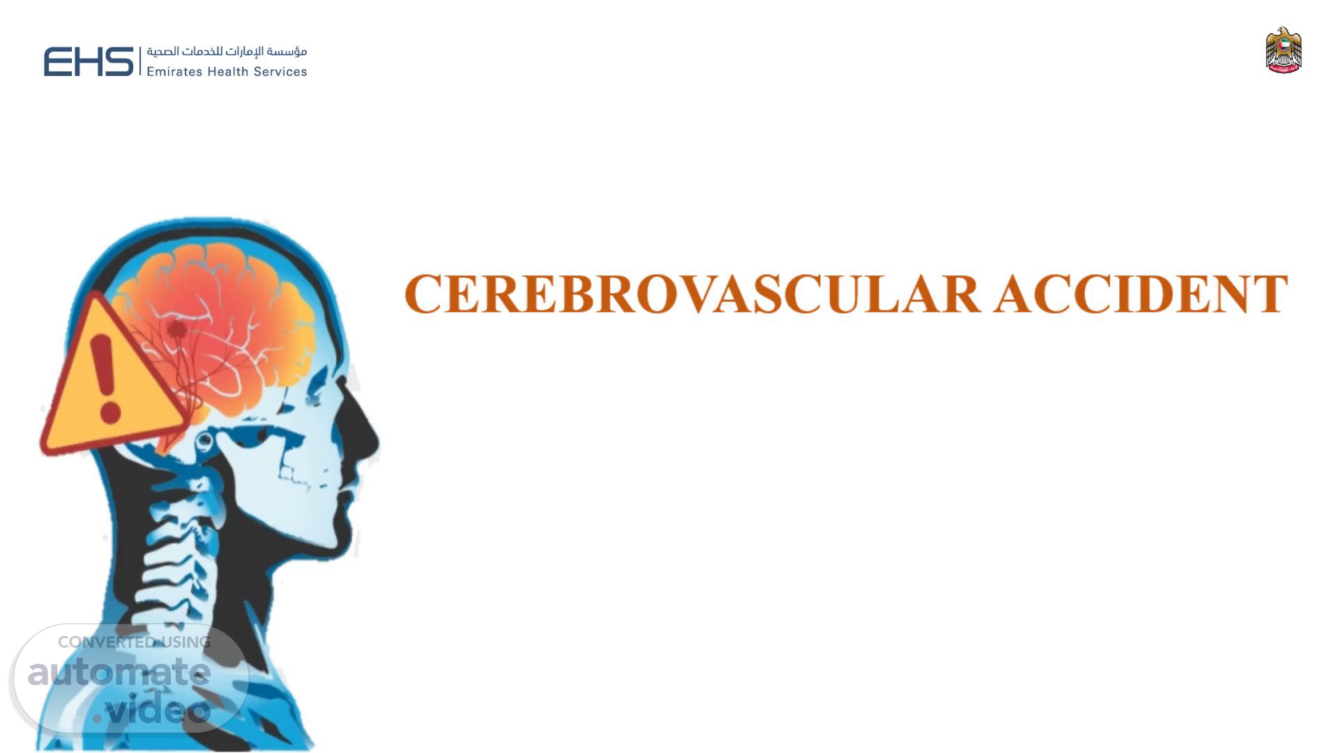
[Audio] CEREBROVASCULAR ACCIDENT Presenter: Hinisha Kumari I, CRN Masafi Hospital.
Scene 2 (13s)
[Audio] Learning Objectives By the end of the session, the learner will be able; Describe the anatomy and physiology of Brain Define Cerebrovascular Accident (CVA) Describe etiology and risk factors of CVA Illustrate the pathophysiology of CVA Point out the symptoms of CVA List down the diagnostic evaluation of CVA Explain the treatment modalities of CVA Explain pre and post operative nursing management of CVA.
Scene 3 (43s)
[Audio] ANATOMY OF THE BRAIN The human brain is the central organ of the human nervous system, and with the spinal cord makes up the central nervous system Brain is composed of three parts; Cerebrum Cerebellum Brainstem..
Scene 4 (59s)
[Audio] PHYSIOLOGY OF THE BRAIN The Brain consists of four main structures: the Cerebrum, the Cerebellum, the Pons, and the Medulla. The Cerebrum is the upper part of the brain and is arranged in two hemispheres called cerebral hemispheres. The cerebrum is thought to control conscious mental processes. The outer layer of the cerebrum is called gray matter, the inner portion, white matter. The cerebral hemispheres are divided into four sections or lobes: frontal lobe, responsible for thinking, making judgments, planning, decision-making and conscious emotions Parietal Lobe, mainly associated with spatial computation, body orientation and attention Temporal Lobe, concerned with hearing, language and memory Occipital Lobe, mainly dedicated to visual processing..
Scene 5 (1m 53s)
[Audio] The Cerebellum is the part of the brain located between the brain stem and the back of the cerebrum. The cerebellum controls muscle coordination and maintains bodily equilibrium. The Pons is in front of the cerebellum and coordinates the activities of the cerebrum and the cerebellum by receiving and sending impulses from them to the spinal cord. The Medulla is part of the brainstem situated between the pons and the spinal cord and it controls breathing, heartbeat, and vomiting. The Meninges consist of three membranes which cover the brain and spinal cord including the dura mater, the arachnoid membrane and the pia mater..
Scene 6 (2m 33s)
[Audio] CEREBROVASCULAR ACCIDENT A stroke, also referred to as a cerebral vascular accident (CVA) or a brain attack, is an interruption in the flow of blood to cells in the brain Also called as ‘Brain attack’, Cerebral hemorrhage, Cerebral infarction, Ischemic stroke or Stroke.
Scene 7 (2m 55s)
[Audio] PREVALENCE OF CVA It is estimated that CVA is the second leading cause of disability in the UAE, with 10,000 to 12,000 stroke patients a year. This means every hour, one person gets a stroke in the UAE. 50% of the stroke patients in the UAE are below the age of 45, as compared to the global average, where 80 per cent of stroke patients are above the age of 65 In the UAE, 18 to 20 percent of the population is obese, and 20% of population are diabetic. Sedentary lifestyle, diabetes, obesity, dependence on fatty foods and a diet high in salts..
Scene 8 (3m 39s)
[Audio] ETIOLOGY OF CVA Cerebral thrombosis – a blood clot or plaque blocks an artery that supplies a vital brain center Cerebral hemorrhage or aneurysm – an artery in the brain bursts, weakens the aneurysm wall; severe rise in BP causing hemorrhage and ischemia Cerebral embolism – a blood clot breaks off from a thrombus elsewhere in the body, lodges in a blood vessel in the brain and shuts off blood supply to that part of the brain..
Scene 9 (4m 9s)
[Audio] RISK FACTORS OF CVA Being over age 55 Being an African-American authenticity Having diabetes Having a family history of stroke.
Scene 10 (4m 21s)
[Audio] . Image result for cva pathophysiology.
Scene 11 (4m 28s)
[Audio] Part 11: Adult Stroke | Circulation. Part 11: Adult Stroke | Circulation.
Scene 12 (4m 34s)
[Audio] TYPES OF STROKE Ischemic stroke - when arteries are blocked by blood clots or by gradual build up of plaque and other fatty deposits. Almost 85% of strokes are ischemic. Hemorrhagic stroke – when a blood vessel in the brain breaks leaking blood into the brain. About 15% of all strokes but responsible for 30% of stroke deaths..
Scene 13 (4m 57s)
[Audio] STAGES OF STROKE Transient ischemic attack (TIA) – sudden and short-lived attack Reversible ischemic neurologic deficit (RIND) similar to TIA, but symptoms can last up to a week Stroke in evolution (SIE) - gradual worsening of symptoms of brain ischemia Completed stroke (CS) – symptoms of stroke stable over a period and rehab can begin.
Scene 14 (5m 27s)
[Audio] CLINICAL SIGNS In Embolism Usually occurs without warning Client often with history of cardiovascular disease In Thrombosis Dizzy spells or sudden memory loss No pain, and client may ignore symptoms In Cerebral Hemorrhage May have warning like dizziness and ringing in the ears (tinnitus) Violent headache, with nausea and vomiting..
Scene 15 (5m 50s)
[Audio] Sudden-onset CVA Usually most severe Loss of consciousness Face becomes red Breathing is noisy and strained Pulse is slow but full and bounding Elevated BP May be in a deep coma.
Scene 16 (6m 9s)
[Audio] COMMON STROKE SYMPTOMS Weakness or paralysis Numbness, tingling, decreased sensation Vision changes Speech problems Swallowing difficulties or drooling Loss of memory Vertigo (spinning sensation) Loss of balance and coordination Personality changes Mood changes (depression, apathy) Drowsiness, lethargy, or loss of consciousness Uncontrollable eye movements or eyelid drooping.
Scene 17 (6m 41s)
[Audio] DIAGNOSIS Early Diagnosis and Treatment is the key to prevent further complications . No single test can diagnose . Most doctors use a combination of the following methods to diagnose the disease and rule out other conditions. Medical History Physical Examination Lab Tests Radiology.
Scene 18 (7m 2s)
[Audio] MEDICAL HISTORY The doctor begins by asking the patient to describe the symptoms, and when and how the condition started, as well as how the symptoms have changed over time. The doctor will also ask about any other medical problems the patient and close family members have and about any medications the patient is taking. Accurate answers to these questions can help the doctor make a diagnosis and understand the impact the disease has on patient’s life. C:\Users\Pratheesh.Pillai\Desktop\figure1.webp.
Scene 19 (7m 39s)
[Audio] PHYSICAL EXAMINATION Doctor will be looking for common features in the CVA ACT FAST F = Face Ask the person to smile. Does one side of the face droop? A = Arms Ask the person to raise both arms. Does one arm drift downward? S =Speech Ask the person to repeat a simple sentence. Does the speech sound slurred or strange? T = Time Call 999 immediately!.
Scene 20 (8m 10s)
[Audio] BLOOD INVESTIGATIONS A doctor may perform blood tests to find out how quickly the clots occur, the levels of particular substances in the blood, including clotting factors and whether or not an infection is present. Lumbar puncture Used to assess presence of blood in the CSF.
Scene 21 (8m 34s)
[Audio] RADIOLOGICAL INVESTIGATIONS Magnetic resonance imaging (MRI) and/or computed tomography (CT) imaging, computed axial tomography (CAT) scan: Used to identify edema, ischemia and necrosis Magnetic resonance angiography (MRA) or cerebral Angiography: To identify presence of cerebral hemorrhage, abnormal vessel structures, vessel ruptures, and regional perfusion of blood flow in the brain.
Scene 22 (9m 7s)
[Audio] How is CVA Treated? THROMBOLYTIC THERAPY Anticoagulants: Sodium heparin, warfarin (Coumadin) Antiplatelets: Ticlopidine (Ticlid), clopidogre (Plavix) Antiepileptic medications: Phenytoin (Dilantin), gabapentin (Neurontin) Tissue plasminogen activator (or Alteplase IV r-tPA) is the only FDA-approved treatment for ischemic strokes. Alteplase IV r-tPA needs to be used within three hours of having a stroke or up to 4.5 hours in certain eligible patients..
Scene 23 (9m 48s)
[Audio] Mechanical removal of clot Endovascular procedure or Thrombectomy: Physical removal of a large blood clot, is another strongly recommended treatment option. To remove the clot, doctors thread a catheter through an artery in the groin up to the blocked artery in the brain. The stent opens and grabs the clot, allowing doctors to then remove the stent with the trapped clot. Special suction tubes may also be used to remove the clot. The procedure should be done within six hours of acute stroke symptoms and only after a patient receives alteplase. Patients must meet certain criteria to be eligible for this procedure..
Scene 24 (10m 29s)
[Audio] MANAGEMENT OF HEMORRHAGIC STROKE A small tube called a catheter can sometimes be threaded up through a major artery in an arm or leg and guided into the brain tissue, allowing the surgeon to use camera technology to help fix the problem. Once the catheter is guided to the source of the bleeding, it deposits a mechanical agent, such as a coil, to prevent further rupture. This type of procedure is endovascular, meaning that the surgeon gains access via the vascular system, making it less invasive than conventional surgical treatment. Sometimes surgery is required to secure a blood vessel at the base of the aneurysm..
Scene 25 (11m 9s)
[Audio] NON-DRUG MANAGEMENT Maintain patent airway. Monitor for changes in the client’s level of consciousness (increased intracranial pressure sign). Elevate the client’s head to reduce ICP and to promote venous drainage. Avoid extreme flexion or extension, maintain the head in a midline neutral position and elevate the head of bed to 30 degrees. Institute seizure precautions. Maintain a non-stimulating environment. Assist with communication skills if the client’s speech is impaired..
Scene 26 (11m 43s)
[Audio] 8. Assist with safe feeding. Assess swallowing reflexes. Thicken liquid to avoid aspiration. Eat in an upright position and swallow with the head and neck flexed slightly forward. Place food in the back of the mouth on the unaffected side. Suction on standby. Maintain skin integrity. Encourage Passive Range of Motion (PROM) every 2 hr to the affected extremities and Active Range of Motion (AROM) every 2 hr to the unaffected extremities.
Scene 27 (12m 17s)
[Audio] 11. Elevate the affected extremities to promote venous return and to reduce swelling. 12.Maintain a safe environment to reduce the risks of falls. 13. Scanning technique (turning head from side to side) when eating and ambulating to compensate for hemianopsia. 14.Provide care to prevent deep-vein thrombosis (sequential compression stockings, frequent position changes, mobilization). 15. Administer medications as prescribed..
Scene 28 (12m 54s)
[Audio] References https://www.mayoclinic.org/diseases-conditions/stroke/symptoms-causes/syc-20350113 https://www.healthline.com/health/cerebrovascular-accident https://bmcnephrol.biomedcentral.com/articles/10.1186/s12882-023-03056-x https://medlineplus.gov/stroke.html.
Scene 29 (13m 15s)
[Audio] Thank You. Thank You. The Head and the Heart: Hemodynamic Derangement in Isolated TBI — Taming the SRU.