Scene 1 (0s)
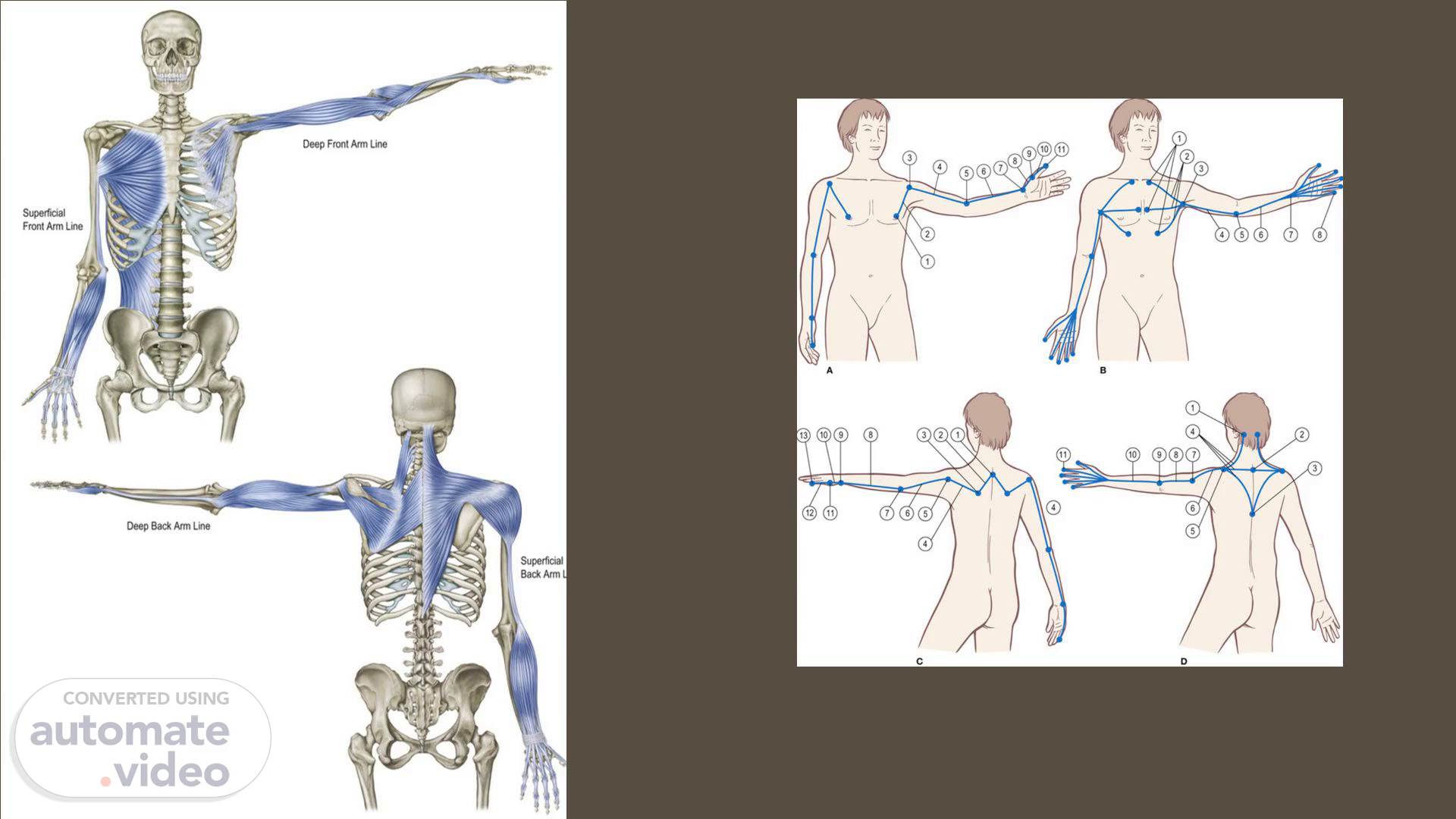
[Audio] From the trunk to the hands, the four Arm Lines run through the four quadrants of the arm. Let's visualize the course of each line. Extend one arm out to the side, palm side forward. The Superficial Front Arm Line initially 'sandwiches' the trunk, spanning from the sternum and the lower back to meet at the top of the upper arm. From there, the myofascial meridian continues in a more 'expected' manner along the front of the arm, all the way to the palm and fingertips. Next, turn the extended arm palm side down, toward the floor. A layer deeper, the Deep Front Arm Line traverses from the chest over the front of the shoulder and anterior edge of the outstretched arm to the thumb. Now turn the arm palm side forward again. The Superficial Back Arm Line starts at the back of the head, neck, and upper back, from where it envelopes the shoulder. The line then stretches along the entirety of the back of the arm to the dorsal side of the hand. Finally, turn the arm palm side down again. A layer deeper, the Deep Back Arm Line runs from the back of the head, neck, and upper back through the shoulder and along the posterior edge of the extended arm to the little finger..
Scene 2 (1m 13s)
[Audio] In the trunk, the SFAL starts with two sizable muscles, which you already met as part of the FLs, the pectoralis major and the latissimus dorsi. Spanning from the rectus sheath, sternum, front ribs, and clavicle to the humerus. The latissimus dorsi begins its embryological life in the front of the trunk before migrating to the back. This connects the SFAL to the back of the pelvis, the thoracolumbar fascia, posterior ribs, and the spine with the front of the upper arm. Living alongside the latissimus dorsi and mirroring its shoulder actions is the teres major. Functionally, it is considered part of the SFAL. In a collaborative effort, pectoralis major, latissimus dorsi, and teres major flex and extend the shoulder, internally rotate the joint, and, through adduction, draw the arm toward the body. When the shoulder is heavily loaded and in demand, these muscles firmly anchor the head of the humerus in the capsule. What is functionally crucial to remember about the thoracolumbar fascia, is that shoulder, arm, and even hand movement influences the state of the lower back. Viewed the other way around, the condition of the lower back affects ease and movement efficiency in the shoulders, arms, and hands. In case you are pondering the part of the thoracolumbar fascia most directly linked with the SFAL, it is the superficial (posterior) layer..
Scene 3 (2m 46s)
[Audio] From the neighboring attachments of the pectoralis major and latissimus dorsi, the SFAL runs towards the elbow with the medial intermuscular septum, a fascial membrane between the flexors and extensors of the upper arm. At the medial epicondyle of the humerus, it reaches into the common flexor tendon, which leads to the superficial forearm flexors. Among these five muscles is the pronator teres, a crossover officially part of the DFAL. The flexor carpi radialis longus, palmaris longus, flexor carpi ulnaris, and the flexor digitorum superficialis make up the rest of the group. When looking to the deep forearm flexors we encounter the flexor digitorum profundus and flexor pollicis longus. Because these two muscles are part of the same fascial complex, they are considered part of the SFAL. Collectively, the superficial and deep flexors bend the wrist and curl all five fingers. In addition, they contribute to dynamic stabilisation of the wrist and hand. The superficial forearm flexors also play a role in dynamic stabilisation of the elbow. Another pathway from the forearm to the palm of the hand is the carpal tunnel, a narrow osteofibrous canal at the inside of the wrist between the flexor retinaculum and the carpal bones..
Scene 4 (4m 6s)
Occipital ridge, nuchal ligament, thoracic spinous processes Trapezius Spine of scapula, acromion, lateral third of clavicle Deltoid Deltoid tubercle of humerus Lateral intermuscular septum Lateral epicondyle of humerus Extensor group Dorsal surface of hand and fingers.
Scene 5 (4m 18s)
1. Pectoralis major, latissimus dorsi, teres major, thoracolumbar fascia:.
Scene 6 (4m 40s)
[Audio] The DFAL starts its journey to the periphery on the ribs and clavicle with the pectoralis minor and subclavius. Both muscles are embedded in the clavipectoral fascia, which lies beneath pectoralis major and spans from clavicle to armpit. The pectoralis minor travels obliquely upward to the coracoid process. When in action on its own, the pectoralis minor draws the shoulder blade forward and down, lifting its inferior angle up and away from its resting place, close to the posterior ribs. Subclavius spans between the first rib and the clavicle. From there, it dynamically stabilises the collar bone relative to the ribcage and relative to the sternum, where we have another frequently overlooked structure, the sternoclavicular joint. Smooth, multidimensional glide in the sternoclavicular joint is essential for easeful shoulder movement..
Scene 7 (5m 33s)
[Audio] From the coracoid process, the DFAL reaches out into the arm with the coracobrachialis and the biceps brachii. The slender coracobrachialis traverses the glenohumeral joint on its way to the humeral shaft. In contrast, the two-headed biceps brachii stretches across the shoulder and the elbow joint. In the forearm, it attaches to the tuberosity of the radius, where it connects with the deep fascia in the forearm. The coracobrachialis competently flexes and adducts the shoulder joint, whereas the biceps brachii contributes only weakly to shoulder flexion. It is, however, adding or taking away from the spaciousness of the shoulder joint (the room between the head of the humerus and the capsule, the glenoid cavity). The big strength of the biceps brachii lies in its capacity to flex the elbow and supinate the forearm. This brings us to the primary flexor of the elbow, the brachialis. From the anterior surface of the humerus, it reaches to the upper part of the ulna..
Scene 8 (6m 38s)
[Audio] Starting where the short head of the biceps brachii left off, the supinator curls around the upper third of the radius. It joins the biceps brachii in forearm supination. Because of its role as a counter-partner of the supinator, the pronator teres can be viewed as a functional crossover from the SFAL. The DFAL continues along the radial periosteum to the styloid process at the distal end of the radius. The fascial connection from the elbow toward the wrist also involves the interosseous membrane between the radius and the ulna. It is a fibrous sheath providing dynamic stabilisation while enabling the bones to smoothly move relative to each other. At the wrist, the DFAL uses the short and sturdy radial collateral ligament to traverse from the lowest aspect of the radius to two carpal bones, the scaphoid and trapezium. The line continues to the thumb, where it completes its journey trunk to periphery with the thenar muscles. These four short muscles include adductor pollicis, abductor pollicis brevis, flexor pollicis brevis, and oppenens pollicis. Collectively, they flex, adduct, abduct, and internally rotate the thumb while contributing to its dynamic stability..
Scene 9 (7m 59s)
Inferior edge of clavicle, 3rd, 4th, 5th ribs Subclavius, pectoralis minor, clavipectoral fascia Coracoid process Coracobrachialis, biceps brachii, brachialis Radial tuberosity Supinator, pronator teres, radial periosteum Styloid process of radius Radial collateral ligament Scaphoid, trapezium Thenar muscles Thumb.
Scene 10 (8m 10s)
Pectoralis minor, subclavius, clavipectoral fascia:.
Scene 11 (8m 40s)
[Audio] The trapezius marks the generous start of the SBAL. Spanning from the occiput along the spine to the lowest thoracic vertebra, it reaches out to the scapula and clavicle. Snug over the shoulder, the deltoid traverses from the shoulder joint outward to the deltoid tuberosity on the humerus. Individual aspects of the trapezius fascially link with individual aspects of the deltoid. With a little bit of imagination, one half of a 'trapezio-deltoid cape' can be visualised. Envisioning the complex functions of these two muscles requires a bit more brain power. The trapezius has the capacity to move the head and the neck or to act on the shoulder girdle, depending on which aspect of the muscle is active and how. The superior or descending part can extend the head and neck, produce ipsilateral side flexion and contralateral rotation, plus deceleration of the same movements in the opposite direction. Contributing to the infamous head-forward position is also within the scope of the upper trapezius. When directing our attention to the shoulder girdle, we see a lift in the clavicle and a drag of the scapula upward and inward. The middle aspect of the trapezius draws the shoulder blade toward the spine. The inferior or ascending portion slides the scapula downward and inward while turning the glenoid cavity upward. You can envision how the upper and lower trapezius function as counter-partnering aspects of the same muscle. Looking at the complete array of functions, the role of trapezius in dynamic stabilisation of the shoulder girdle is evident. So is the necessity to keep its individual parts in balance with each other and with other myofascial units acting on the shoulder blade. With the anterior or clavicular portion in action, the deltoid flexes and internally rotates the shoulder joint. The medial or acromial aspect abducts the arm. The posterior or spinal part produces shoulder extension and external rotation. Together, the anterior and posterior portions 'neutralise' or, better phrased, dynamically balance each other when raising the arm sideways..
Scene 12 (10m 56s)
[Audio] From where the deltoid left off, the lateral intermuscular septum, a fascial wall separating the extensors and flexors of the upper arm, spans to the lateral common extensor tendon, which reaches into the extensors of the wrist and hand. The superficial forearm extensors encompass anconeus, brachioradialis, extensor carpi ulnaris, extensor carpi radialis longus, extensor carpi radialis brevis, extensor digitorum, and extensor digiti minimi. Turning our attention to the deep forearm extensors brings us to the extensor pollicis longus, extensor pollicis brevis, abductor pollicis longus, and extensor indicis. Another member of this group is the supinator, a crossover from the DFAL. Collectively, the superficial and deep extensors extend the wrist and fingers while contributing to dynamic stabilisation all around. The superficial forearm extensors play a role in dynamic stabilisation of the elbow..
Scene 13 (12m 1s)
Occipital ridge, nuchal ligament, thoracic spinous processes Trapezius Spine of scapula, acromion, lateral third of clavicle Deltoid Deltoid tubercle of humerus Lateral intermuscular septum Lateral epicondyle of humerus Extensor group Dorsal surface of hand and fingers.
Scene 14 (12m 22s)
Trapezius, deltoid:. Scapula elevation, adduction, depression, and internal rotation (glenoid cavity turns upward)a Multidimensional dynamic scapula stability Shoulder joint flexion and extension, internal and external rotation, and abduction Weight-bearing strength when supporting the body on hands and arms.
Scene 15 (12m 43s)
[Audio] In the DBAL, we will identify two related myofascial tracks travelling from the spine to the humerus. The first one starts on the back of the head with rectus capitis lateralis, a small muscle connecting the underside of the occiput with the first vertebra. At the atlas, the much more prominent levator scapulae joins the line. Technically a back muscle but functionally considered a scapula stabiliser, it reaches from the cervical spine down to the most upper portion of the medial border of the scapula. Following the fibers leads us into the supraspinous fossa on the top of the shoulder blade and into the supraspinatus, one of the four rotator cuff muscles. The supraspinatus connects to the greater tubercle of the humerus. Functionally, the levator scapula acts on the cervical spine and the shoulder blade. When the shoulder blade is stabilized, the levator scapula laterally flexes the neck and produces extension. In a well-disposed way, you could say it prevents the upper neck from shifting sideways and forward. With the neck securely stabilised, the levator scapulae draws the shoulder blade upward and inward, turning the glenoid cavity downward. Although the supraspinatus contributes to shoulder abduction, it first and foremost acts as a dynamic stabiliser of the shoulder joint. Turning the attention back to the spine, we are going to follow the other track of the DBAL. From the lowest cervical and upper thoracic spine, the rhomboid minor and rhomboid major traverse to the medial border of the scapula, where we encounter a switch. With the SPL, we follow the fabric into the serratus anterior (remember the 'rhombo-serratus muscle'). With the DBAL, we reach into the infraspinatus, another member of the rotator cuff. On its way to the greater tubercle of the humerus, the infraspinatus picks up the teres minor, a muscle that is also part of the rotator cuff group. In the realm of movement, the rhomboids elevate and adduct the scapula while turning the glenoid cavity downward. The infraspinatus and teres minor externally rotate the shoulder and, most importantly, act as dynamic balancers and stabilisers of the joint. What about the fourth rotator cuff muscle, the subscapularis, you might wonder. Located on the underside of the shoulder blade, it is fascially tensioned when the rhomboid tugs on the bone. In addition, it clearly plays a crucial role in dynamic shoulder stabilisation, which makes its integration into the DBAL easy to justify. Complementing the other three rotator cuff muscles, the subscapularis internally rotates the shoulder joint..
Scene 16 (15m 29s)
[Audio] From the top of the lateral side of the scapula, near the teres minor and the shaft of the humerus, close to where the rotator cuff muscles attach, the triceps brachii spans the upper arm. It crosses the elbow joint to link with the olecranon of the ulna and the antebrachial fascia. In its movement scope are shoulder extension and adduction, and most importantly, elbow extension nd deceleration of flexion. With the periosteum of the ulna and adjacent fascial layers, the DBAL travels to the styloid process on the outside of the wrist. Ligaments of the wrist, especially the stabilising ulnar collateral ligament, serve as pathways across the midcarpal joint. Along the way, the ulnar collateral ligament contacts two carpal bones, the triquetrum and hamate. With the hypothenar muscles, the DBAL completes its journey on the flexor digiti minimi, opponens digiti minimi, and palmaris brevis encompass flexing and abducting the little finger while providing dynamic stabilisation..
Scene 17 (16m 37s)
Inferior surface of occiput, transverse processes C1-C4 and spinous processes C7-T5 Rectus capitis lateralis, levator scapulae, rhomboid minor, rhomboid major Medial border of scapula Rotator cuff muscles (supraspinatus, infraspinatus, teres minor, subscapularis) Head of humerus Triceps brachii Olecranon of ulna Fascia along ulnar periosteum Styloid process of ulna Ulnar collateral ligament Triquetrum, hamate Hypothenar muscles Little finger.
Scene 18 (16m 49s)
Rectus capitis lateralis, levator scapulae, rhomboids, rotator cuff muscles:.
Scene 19 (17m 15s)
[Audio] This is a table of mentioned crossovers. Remember, a crossover is a myofascial 'bridge' between two or more ALs. Because force is transmitted via this bridge, a functional connection is created. Meaning activation of one line immediately evokes change in the other(s)..