Scene 1 (0s)
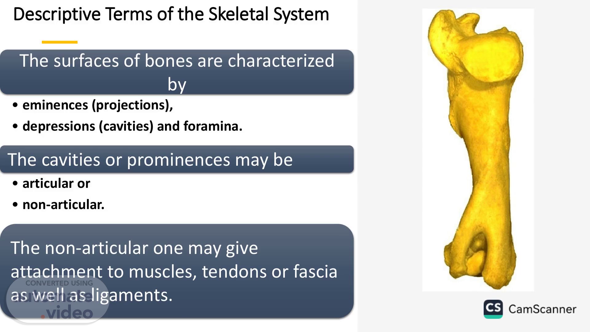
[Virtual Presenter] Descriptive Terms of the Skeletal System The surfaces of bones are characterized by eminences (projections), depressions (cavities) and foramina. The cavities or prominences may be articular or non-articular. The non-articular one may give attachment to muscles, tendons or fascia as well as ligaments..
Scene 2 (26s)
[Audio] Non articular projections Process is a general term of a prominence which is nonarticular (e.g. Spinous Process of the Scapula). Tuberosity is a large rounded non-articular projection (e.g. Metacarpal Tuberosity). Tubercle is similar to the above but smaller. Trochanter is a non-articular prominence (e.g. Trochanters of the femur)..
Scene 3 (51s)
[Audio] Non articular projections (con.) Spine or spinous process is a pointed projection (e.g. Dorsal & Ventral spines of vertebrae or soine of scapula). Crest is a sharp ridge. Line is a very small ridge (e.g. muscular lines of the subscapular fossa). Epicondyle is a non-articular projection in connection with the condyle (e.g. Epicondyle of the femur) Hamulus is hook like projection Malleolus is round projection (e.g. medial and lateral malleolus of tibia).
Scene 4 (1m 35s)
[Audio] Articular projections Head is a rounded articular enlargement placed at the proximal end of the bone, and usually medial in position (e.g. Head of Humerus & Femur). Condyle is an articular eminence, somewhat cylindrical in shape (e.g. Condyles of the femur Trochlea Is a pulley like articular mass (e.g. Trochlea of the femur) Facet is small flat articular surface (e.g. costal facet of vertebrae).
Scene 5 (2m 5s)
[Audio] Non articular depressions Fossa is large non articular depression of the bones (e.g. supraspinous fossa of scapula) Fovea is small non articular depression (e.g. fovea capitis of head of femur) Groove (sulcus) is elongated usually deep (e.g. musculospiral grove of humerus) Foramen is circumscribed hole or perforation of the bone ( nutrient foramen) Fissure is liner defect of the bone or between two bones Canal is tunnel through one or more bone ( e.g. vertebral canal) Meatus is short canal (e.g. external acuastic meatus of ear) Hiatus : canal ( aortic hiatus of diaphragm) Alveolus is the tooth socket.
Scene 6 (3m 0s)
[Audio] Articular depressions Glenoid cavity is a shallow articular cavity (e.g. glenoid cavity of the scapula). Acetabulum or cotyloid cavity is a deep articular cavity (e.g. Acetabulum of the pelvic bone). Notch may be articular depression of bone (semilunar notch of ulna). It usually used also to describe non articular depression.
Scene 7 (3m 33s)
[Audio] Skeleton of horse. Skeleton of horse.
Scene 8 (3m 39s)
Parietal b. Temporal b. Frontal b. Zygomatic b. Lacrimal b. Nasal b. Infraorbital foramen Maxilla Incisive b. External acoustic meatus Occipital b. Atlanto-occipital joint Atlas (Cl) Atlantoaxial joint Axis (C2) Scapular cartilage Scapular spine Wing Of ilium Sacral tuber Scapula Costovertebral joint T18 Mandible L6 Ll 18th rib Facial crest 1st rib Mental foramen Supraglenoid tubercle Greater tubercle Humerus Deltoid tuberosity Coxal tuber Body of ilium Pubis Sacrum Caudal vertebrae Ischiatic tuber Ischium Greater trochanter Femur Patella Olecranon tuber Ulna Proximal phalanx (long pastern b.) Middle phalanx (short pastern b.) Sternum Lateral epicondyle Radius Accessory carpal b. Intermediate carpal b. Ulnar carpal b. 3rd carpal b. 4th carpal b. 3rd metacarpal b. (cannon bone) 4th metacarpal b. (lateral splint b.) Lateral proximal sesamoid b. Distal phalanx (coffin bone) Xiphoid process Sternocostal joint Talu Central tarsal b. 3rd tarsal b. 3rd metatarsal b. Central tarsal b. Lateral epicondyle Fibula Tibia Calcaneal tuber Calcaneus Talus 4th tarsal b. 4th metatarsal b. 3rd carpal b. Radial carpal b. 2nd carpal b. 2nd metacarpal b. (medial splint b.) Medial proximal sesamoid b. Distal sesamoid b. 3rd tarsal b. (lateral splint b.) fused 1st & 2nd tarsal b. 2nd metatarsal b. (medial splint b.) cs CamScanner.
Scene 9 (4m 20s)
Horse and Human Common Parts T" and Fua R.rs Metacypal bues paWa Raags Aun HUMAN tu•rMus Rivis Scania Wrist Kne cs CamScanner.
Scene 10 (4m 27s)
[Audio] ForelimbOrThoraciclimb. Fore limb Or Thoracic limb.
Scene 11 (4m 33s)
[Audio] The bones of the thoracic limb (forelimb) of domestic animals are divided into 4 regions. shoulder girdle A.The shoulder girdle: When fully developed it consists of 1 The scapula (shoulder blade): 2- The coracoid: (In domestic animal as a coracoid The Arm process). 3- The clavicle (clavicula) it is absent in domestic The Forearm mammals or it is a small rudiment embedded in the brachiocephalic muscle (dog & cat). Therefore, there is no articulation between the forelimb and axial skeleton. B. The Arm (Brachium): Humerus. C. The Forearm (Antibrachium): The Manus 1- The radius. 2- The ulna..
Scene 12 (5m 26s)
[Audio] shoulder girdle D. The Manus: It is similar to the hand in man, it consisted of 3 subdivisions: 1. The Carpus (carpal bones) or Wrist: In two rows, proximal and distal, each of which typically contains four bones The Arm from 6-8 in the different domestic animals. Only pig and man have the 8 carpal bones. 2. The Metacarpus: The Forearm typically (5) metacarpal bones (I-V) one for each digit. In the horse the first and 5th are absent, 2nd and 4th are reduced and the 3rd is the largest well developed metacarpal bone Dog has 5 metacarpal bones, the first metacarpal is much reduced. The Manus.
Scene 13 (6m 23s)
[Audio] 3. The Digits: These are homologues to the fingers of man and are typically 5 in number. Only dog has 5 digits while in other domestic animals the number of digits is varied. Each fully developed digit consists of 3 phalanges; the Proximal (First) Phalanx, the Middle (Second) Phalanx and the Distal (Third) Phalanx. Each digit has a 2 proximal and one distal sesamoid bones (distal one is absent in dog and camel).
Scene 14 (6m 59s)
[Audio] Comparative anatomy of scapula The scapula is triangular in outline lies against the cranial part of the lateral thoracic Directed cranioventral direction. It is linked to the trunk by muscles (synsarcosis) without forming a true articulation. It is triangular in outline and has 2 surfaces, 3 borders and 3 angles..
Scene 15 (7m 24s)
capu a o orse Lateral surface Scapular cartilage Cranial angle Infraspinous fossa Supraspinous fossa Tuberosity of scapular spine Scapular spine Cranial bor&r Scapular notch Supraglenoid tubercle Glenoid cavity Caudal angle Caudal border Medial surface Scapular cartilage Cranial angle Roughened area ace of attachment ventral serrate muscle) Subscapular fossa Cranial border Scapular notch Coracoid process Glenoid cavity cs.
Scene 16 (7m 35s)
[Audio] Thelateralsurface: 1.itisdividedspineofthescapula(spinascapulae)intothesmallercranialsupraspinousfossaandthelargerinfraspinousfossacaudally. Thespineextendsformthevertebralbordertotheneckofthebone,whereitdisappears.supraspinousfossatuberspinae Atitsmiddle,thereisaroughprominenceknownasinfraspinousfossaspinethetuberosityofspineofscapula(tuberspinaescapulae).
Scene 17 (8m 17s)
[Audio] The Medial (Costal) surface: It has shallow subscapular fossa, . Proximally in both side of fossa there are two triangular rough areas (facies serrata), Vertebral border facies serrata cranial border facies serrata 1. The cranial border: It is convex and rough subscapular fossa above, concave and smooth below 2. Caudal border: It is thick and rough in its proximal third thin in its middle, and thick below. caudal border 3. The vertebral border: It carries the scapular cartilage.
Scene 18 (9m 0s)
[Audio] 1. The cranial angle: it is thin and lies between the cranial and vertebral borders. Cranial angle 2. The caudal angle: it is rough and thick and Caudal angle lies between vertebral and caudal borders supraspinous fossa tuber spinae 3. Articular or glenoid angle: it separated infraspinous fossa spine from the body of bone by neck and it has a. Glenoid cavity: b. glenoid rim and notch c. supraglenoid tubercle or tuber scapulae d. The coracoid process Glenoid angle.
Scene 19 (9m 42s)
capu a o orse Lateral surface Medial surface Scapular cartilage Cranial angle Roughened area lace of attachment Er ventral serrate muscle) Subscapular fossa Cranial border Scapular notch Coracoid process Glenoid cavity Supraglenoid tubercle Coracoid process Glenoid cavity Scapular cartilage Cranial angle Infraspinous fossa Supraspinous fossa Tuberosity of scapular spine Scapular spine Cranial border Scapular notch Supraglenoid tubercle Glenoid cavity Caudal angle Caudal border Glenoid notch cs CamScanner.
Scene 20 (9m 54s)
[Audio] Scapulaofdog 1.Thelateralsurfaceisdividedbythespineofthescapulainto2nearlyequalfossae 2.Spineextendsandprojectasacromionprocessandreachnearlytothelevelofglenoid 3.Theacromionprocessofdogusuallyknownashamatusprocess.Tuberspinaeisabsent 4.Cranialborderisthinandhavescapularnotchdistally 5.Glenoidcavityisshallowandcoracoidprocessissmall.
Scene 21 (10m 38s)
[Audio] Scapula of ox 1. The scapula of ox is more triangular, wide at its vertebral border and narrows distally 2. The spine is situated further cranial than that of horse. It ends by pointed acromion process, with no distinct tuber spinae. 3. Supraspinous fossa is narrow proximally and disappear distally 4. Glenoid is nearly circular without notch 5. Subscapular fossa is shallow and facies serrate is not distinct..
Scene 22 (11m 21s)
[Audio] Sheep Scapula of camel 1. The spine is thick and form projected acromion process that reaching the level of glenoid 2. Infraspinous fossa is slightly larger than supaspinous fossa (5/4) 3. Glenoid is shallow circular and the notch is absent. 4. Nutrient foramen is situated in the distal third of the infraspinous fossa. 5. The tuber scapulae is very large. The coracoid process is.