Scene 1 (0s)
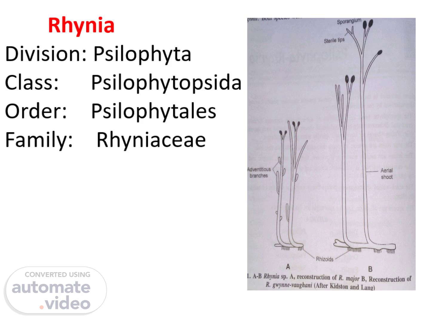
Rhynia Division: Psilophyta Class: Psilophytopsida Order: Psilophytales Family: Rhyniaceae.
Scene 2 (15s)
Occurrence: fossil Rhynia was discovered from lower Devonian red sandstone cherts of village Rhynie in Scotland by Kidson & Lang 1917. Plant was named after the locality Rhynia village. Rhynia has 2- well defined species Rhynia gwynne vaughanii Rhynia major Grew in marshy environment Plant body was sporophyte.
Scene 3 (30s)
[image] General Features UCIassification Class: Psilophytopsida — Order: Psilophytales - Family: Rhyniaceae - • Discovered by: Robert Kidston & William H. Lang (1917), R. major (1920) • Rhynia gwynne-vaughanii • Location: Rhynie chert in Aberdeenshire, Scotland • Named in honor of botanist Helen Gwynne- Vaughan • Preserved in Hard siliceous rocks of Lower Devonian age. • Evidenced suggest that they grow near volcanoes in habitats saturated with acid water from hot springs. 'ants exist earlier tips.
Scene 4 (50s)
External features: Rootless, dichotomously branched creeping rhizome with cylindrical, erect leafless dichotomously branched shoots. Some of them terminated into sporangia. Rhizome had rhizoids that helped in attachment & absorption of water.
Scene 5 (1m 2s)
Rhynia gwynne vaughanii: small plant with 17-20 cm in height dichotomously branched stem arising from subterranean horizontal rhizome Rhizome lacked roots. Aerial stem terminated into fusiform sporangia & lacked leaves. Stem possessed hemispherical projections & considered to be adventitious branches. Rhynia major: stout plant attaining the height of 51cm. Both species are similar except hemispherical projections..
Scene 6 (1m 20s)
Internal structure: stem: Epidermis: single layer of cells covered externally by thick layer of cuticle & interrupted by stomata. Each stomata consisted of 2-guard cells surrounded by many subsidiary cells. Cortex: differentiated into outer & inner cortex. Outer cortex: comprised of a narrow band of 1-4 layer of large polygonal compactly arranged cells without any intercellular spaces. Inner cortex: consisted of large number of circular cells with intercellular spaces that were connected to outside through the stomata present in the epidermis..
Scene 7 (1m 44s)
[image] Internal Structure of Stem Central cylinder: Protostele present in central part, amphigribal (xylem surrounded by phloem). Xylem- only with 09 annular or spiral thickening. Phloem- 4-5 layered thin elongated cells. Endodermis & pericycle however absent. walled were, Stoma —Cuticle Epidermis Outer cortex Inner cortex Xylem Phloem.
Scene 8 (1m 58s)
Cuticle Epidermis Outer a»rtex Inner cortex Phloem M etaxyiern (A) 8 Protoxylem Stoma Cuticle Epidermis Outer cortex Inner cortex Xylem Phloem o (B) Fig. 3.2. A-B A, T.S. of aerial shoot of Rhynia moor; B, Rhynia gwynne-vaughani, T.S. of an aerial branch..
Scene 9 (2m 13s)
Stele: Protostele Consisted of central core of xylem surrounded by phloem was present in the centre of the stem. Xylem showed tracheids with spiral or annular thickenings. Phloem was made up of 4-6 layers of thin walled elongated cells. Endodermis & pericycle were absent..
Scene 10 (2m 28s)
Reproduction: gametophytes were dioecious with antheridia & archegonia borne on different axis. Sporangia: oval or cylindrical sporangia were Borne singly on apex of certain branches of aerial stem. Sporangia of R. major were 12 mm in length & 4mm in diameter while in R. gwynne vaughanii Were 3mm in length & 1-1.5 mm in diameter. Both species had structurally similar sporangia that were cylindrical in shape & bounded by multilayered thick wall..
Scene 11 (2m 49s)
Archegonium Fig. 3.4. Archegonium of Rhynia gwynne-vaughani.
Scene 12 (2m 57s)
[image] Sporangia: Reproductive structure • Born singly on aerial branches, oval or slightly cylindrical. • R. myor: 12 mm long & up to 4 mm in breadth. • R. gwynne-vaughanii: comparatively small. • Sporangium surrounded by multilayered jacket. • Outer wall- made of cells having thick cuticle. • Inner wall- 2-3 layers of thin walled palisade like cells (formed a nutritive layer like tapetum). • Numerous spore tetrad in sporangial cavity. • Spores same type (homosporous). Arrangement shows that they formed by reduction division (meiosis). • Dispersal by decay of sporangia (no specialized mechanism of dehiscence of sporangia). Tapetum.
Scene 13 (3m 23s)
Jacket Tapetirn Spores Aerial shoot Jacket Tapetum Spores eet9 fig. 3.3. A-D Rhynia gwynne•vaughani, A & B, Longitudinal section of C, Spoore Tetrad; D, asin le s.
Scene 14 (3m 33s)
Outer sporangial wall was made up of thick walled vertically elongated cells surrounded by a strong cuticle. Innermost layer was composed of thin walled round cells that formed nutritive layer like tapetum. Numerous spore tetrads of same size (homosporous) were present within the sporangium. Presence of spore tetrad indicates the occurrence of meiosis in the life history..