Scene 1 (0s)
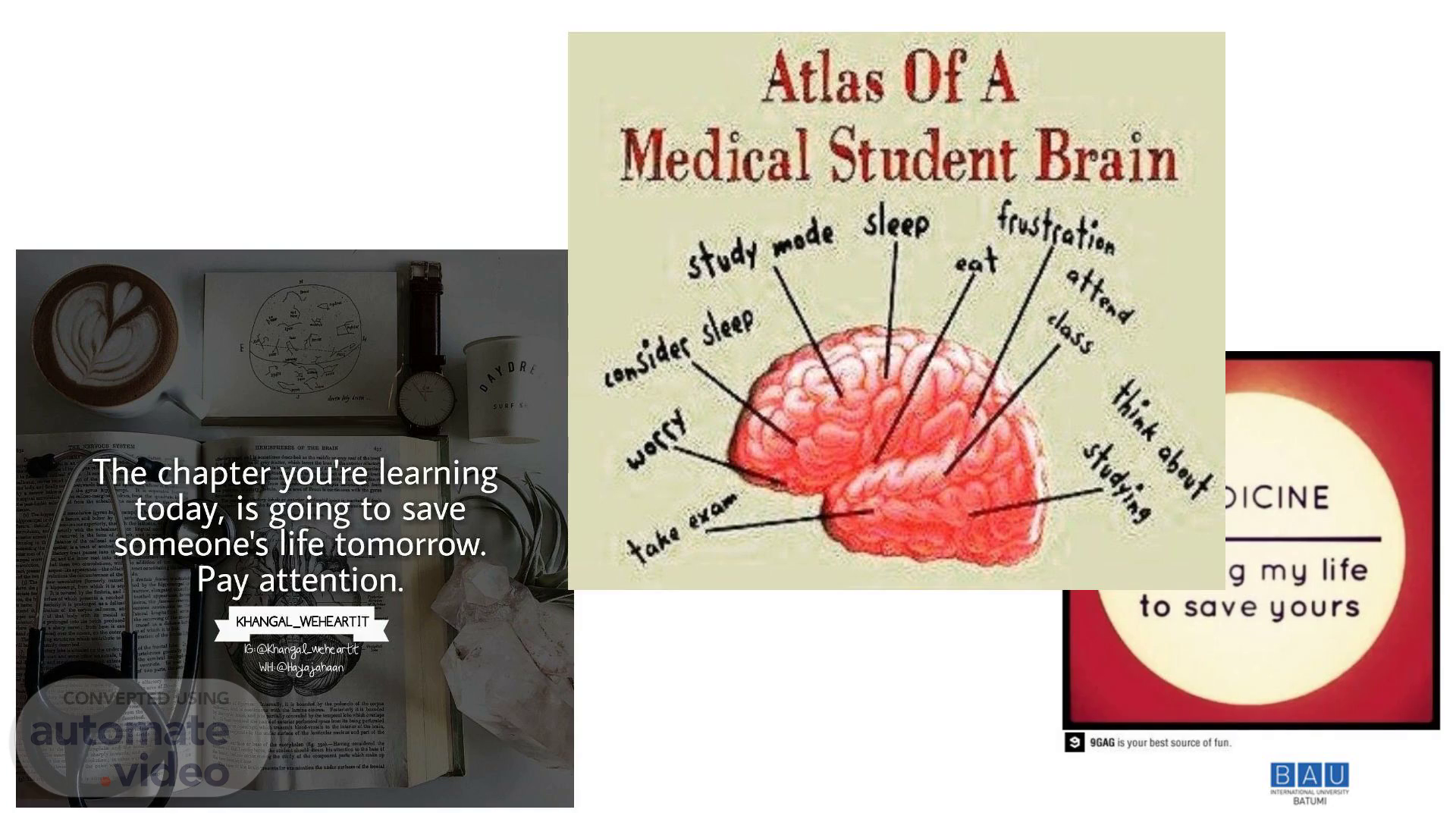
hechapter you're learning today, is going to save someone's life tomorrow. Pay attention. KHANGAL_WEÆARTIT qrtt.
Scene 2 (15s)
Motor & Sensory Functions. Natia Kharati Batumi, 2025.
Scene 3 (24s)
[Audio] Our eyes are responsible for our ability to see, but Dr. Oliver Sacks reminds us that our brains also play a crucial role in this process. His quote highlights the complex relationship between our visual perception and the workings of our brain. By exploring this idea, we can gain a deeper understanding of how our senses interact with our cognitive abilities..
Scene 4 (46s)
[Audio] The motor homunculus is a representation of the body parts corresponding to specific areas of the brain. The precentral gyrus controls various movements, ranging from simple actions like finger movement to complex tasks such as playing a musical instrument. This mapping illustrates the close connection between the brain and physical abilities..
Scene 5 (1m 8s)
[Audio] The motor function is controlled by two main groups of descending tracts, originating from the brain and descending into the spinal cord. These tracts are responsible for various motor activities throughout the body. The first group is the pyramidal tract, also known as the corticospinal tract, which plays a crucial role in voluntary movements such as walking, talking, and writing. The second group is the extra-pyramidal tract, which includes tracts such as the rubrospinal, vestibulospinal, and tectospinal tracts, among others. These tracts are involved in involuntary movements like reflexes, as well as regulating posture and balance..
Scene 6 (1m 47s)
[Audio] The pyramidal pathway, also known as the corticospinal tract, is responsible for transmitting voluntary motor signals from the cerebral cortex to the spinal cord. This pathway plays a crucial role in controlling fine motor movements, such as those required for writing or playing musical instruments. The cortico-spinal tract is composed of fibers that originate in the precentral gyrus and project to the anterior horn cells of the spinal cord. These fibers are responsible for transmitting information regarding muscle contraction and relaxation, allowing for precise control over movement. In addition to the cortico-spinal tract, there are other extrapyramidal pathways that contribute to motor function, including the rubrospinal, vestibulospinal, reticulospinal, tectospinal, and olivospinal tracts. These pathways transmit information from various parts of the brain, such as the red nucleus, vestibular nuclei, and reticular formation, to the spinal cord, influencing motor activity..
Scene 7 (2m 43s)
[Audio] The extrapiramidal pathway plays a crucial role in controlling voluntary movements, particularly fine motor skills. This pathway involves direct connections between the cerebral cortex and distal muscles, allowing for precise control over movements such as clapping or grasping. In contrast, involuntary functions like postural movement, proprioception, and reflexes rely on indirect connections between the cerebral cortex and proximal/axial muscles. These indirect connections result in characteristic patterns of movement, such as cogwheel rigidity or shuffling gait. Furthermore, the presence or absence of superficial reflexes can also provide valuable information about the integrity of this pathway..
Scene 8 (3m 29s)
[Audio] Weakness refers to a reduction in the power that can be exerted by one or more muscles. It's essential to distinguish between weakness and increased fatigability or limitations caused by pain or joint stiffness, which may be mistakenly attributed to weakness by patients. Furthermore, increased fatigability is characterized by the inability to maintain normal activities, while bradykinesia, or slow movement, might also be misinterpreted as weakness. Additionally, apraxia, a condition affecting the planning and initiation of movements, can sometimes be mistaken for weakness..
Scene 9 (4m 3s)
[Audio] Paralysis and paresis are two terms used to describe muscle weakness. While paralysis implies a complete loss of muscle function, paresis suggests a milder form of weakness. The prefixes "hemi-", "para-", and "quadri-" refer to different parts of the body, such as one half, both legs, and all four limbs respectively. On the other hand, the suffix "-plegia" indicates severe weakness or paralysis. Furthermore, the term "mono-" refers to a single limb..
Scene 10 (4m 33s)
[Audio] Localization begins by identifying weakness. We differentiate between various distributions, including hemiplegia and monoplegia. These conditions are often caused by focal structural lesions, making them easier to localize. Additionally, we consider any associated sensory abnormalities when evaluating these patients..
Scene 11 (4m 56s)
[Audio] Hemiplegia is a condition characterized by partial paralysis or weakness affecting one side of the body. It can result from various types of lesions or injuries to different parts of the nervous system. The condition can be categorized into five types: cerebral lesion, brainstem lesion, spinal lesion, peripheral lesion, and functional hemiplegia. A cerebral lesion typically occurs in the prefrontal cortex or other areas responsible for motor control, resulting in hemiparesis or hemiplegia due to damage to these regions. A brainstem lesion could affect the corticospinal tract, leading to contralateral hemiparesis or hemiplegia. A spinal lesion might cause damage to the lateral corticospinal tract, resulting in ipsilateral hemiparesis or hemiplegia. Peripheral lesions, such as those affecting the brachial plexus, could also lead to hemiparesis or hemiplegia. Functional hemiplegia may arise from psychological or emotional factors, rather than physical damage to the nervous system. Accurate diagnosis and treatment planning are crucial in addressing this complex condition..
Scene 12 (6m 6s)
[Audio] Lesions affecting the motor cortex can result in significant impairments to movement, including weakness and poor control of the affected extremity, which may involve the face, arm, and leg to varying degrees. Depending on the location of the lesion within the cortical homunculus, patients may exhibit incoordination and weakness on the contralateral side, often accompanied by neglect, apraxia, or aphasia. In some cases, sensory loss may also occur in the same distribution..
Scene 13 (6m 36s)
[Audio] The diagram shows the representation of the face, arm, and leg on the hemisphere, with the face and arm being laterally represented, and the leg draped over the top and into the interhemispheric fissure. This information is crucial for understanding motor functions and the brain's relationship with the body, as revealed by Dr. Oliver Sacks' work. Further exploration of key concepts and structures in neuroscience will be continued on subsequent slides..
Scene 14 (7m 5s)
[Audio] Damage to the brainstem can result in various symptoms depending on the location and extent of the injury. A lesion affecting the trigeminal nerve can cause an ipsilateral facial sensory deficit, whereas damage to the cerebellar hemispheres or nuclei may lead to hemiataxia. Furthermore, lesions in different areas of the brainstem can also produce ocular motor weakness, with some locations resulting in an ipsilateral Horner syndrome due to damage to the descending sympathetic tracts..
Scene 15 (7m 35s)
[Audio] The bedside language examination is used to assess cognitive and linguistic abilities in patients with suspected neurological disorders. This type of examination helps identify specific areas of impairment, such as difficulty with spontaneous speech, naming, auditory comprehension, repetition, reading, and writing. The results inform treatment decisions and provide valuable information for diagnosis and prognosis. In cases of patients presenting with dysarthria, apraxia, and dysphonia, a bedside language examination assesses their ability to produce speech sounds, understand spoken language, and read and write effectively. By examining these skills, healthcare providers gain insight into the underlying causes of symptoms and develop targeted interventions to improve communication and quality of life..
Scene 16 (8m 26s)
[Audio] Spinal lesions can result in hemiplegia, sparing the face, but they usually cause bilateral deficits characteristic of myelopathy. Patients with bilateral weakness, bowel or bladder control impairments, and back pain should be considered for a spinal cord lesion, which may include conditions such as spinal hemisection, transverse myelitis, spinal cord compression, and spinal cord infarction..
Scene 17 (8m 52s)
[Audio] When considering a diagnosis of hemiplegia, it's essential to differentiate between central and peripheral lesions. A single peripheral lesion can affect either an arm or a leg, whereas a pair of peripheral lesions affecting both limbs on the same side may occasionally mimic hemiplegia. To accurately diagnose, we need to identify each lesion as being within the distribution of a specific nerve, nerve root, or plexus division. This distinction is crucial because peripheral lesions typically result in depressed tendon reflexes, unlike central lesions where reflexes are often increased..
Scene 18 (9m 25s)
[Audio] A functional lesion can cause improvement in strength with coaching, indicating that the patient has some residual motor function. However, this is often accompanied by give-way weakness, where the patient's muscles may appear strong initially, but then weaken when resistance is applied. Inconsistencies in the examination are also common, such as the ability to perform certain movements, like walking on toes, despite being unable to extend the foot. The Hoover sign is a useful clinical tool for assessing paralysis, as it helps to distinguish between true paralysis and weakness. Finally, patients with a functional lesion may exhibit paralysis in the absence of other signs of motor system dysfunction, such as changes in muscle tone or reflexes..
Scene 19 (10m 10s)
[Audio] Tone refers to the resistance of a muscle to passive stretch. This can manifest in various ways, including spasticity, rigidity, paratonia, and flaccidity. Spasticity is characterized by an increase in tone associated with disease of upper motor neurons. Rigidity is a type of increased tone that is present throughout the range of motion, affecting both flexors and extensors equally. Paratonia, also known as gegenhalten, is another form of increased tone that varies irregularly in response to relaxation, affecting both flexors and extensors equally, typically resulting from disease of the frontal lobes. Weakness, on the other hand, is often accompanied by decreased tone, or normal tone, and is commonly seen in disorders of motor units..
Scene 20 (10m 57s)
[Audio] Upper motor neuron lesions, such as those resulting from stroke or traumatic brain injury, typically elevate muscle stretch reflexes, which can be transiently increased. Cutaneous reflexes may also be affected, often exhibiting an abnormal extensor plantar response, commonly referred to as the Babinski sign. Lower motor neuron lesions, such as those caused by amyotrophic lateral sclerosis, can depress these reflexes due to direct involvement of specific reflex arcs. Myopathic conditions, like muscular dystrophy, tend to preserve muscle stretch reflexes until advanced stages, when they may become attenuated..
Scene 21 (11m 38s)
[Audio] Apraxia, which is also referred to as limb apraxia, is an inability to correctly perform learned skilled movements. This neurological dysfunction affects the execution of movements in the arm or hand. Notably, apraxia is not caused by elementary motor deficits such as weakness or spasticity, nor is it the result of primary sensory deficits or complex visual disorders. Furthermore, apraxia is distinct from abnormal movements or postures, like tremors or dystonic posturing. Moreover, it is not apraxia if the impaired movements result from other cognitive disorders affecting attention, memory, or language comprehension..
Scene 22 (12m 19s)
[Audio] The Examination for Limb Apraxias evaluates the capacity to execute diverse gestures, movements, and tasks, encompassing pantomiming verbal commands, mimicking gestures, sequencing actions, and illustrating conceptual knowledge. This assessment aids in determining whether a patient suffers from apraxia, a neurological condition marked by difficulty executing specific actions despite intact motor function. The examination comprises a series of tests, including transitive and intransitive actions, gesture imitation, sequencing, and utilization of genuine objects. These tests facilitate identifying any discrepancies in the patient's performance, offering valuable insights into their cognitive and motor abilities..
Scene 23 (13m 11s)
[Audio] Ideomotor apraxia is a condition characterized by an inability to correctly perform learned skilled movements. This impairment is often associated with damage to specific regions within the brain. Lesions in the inferior parietal lobe, frontal lobe, and premotor areas, particularly the supplementary motor area, have been linked to ideomotor apraxia. Additionally, subcortical lesions in the basal ganglia, thalamus, and associated white-matter tracts, including the corpus callosum, may also contribute to this condition. Limb apraxias, including ideomotor apraxia, can arise from various central nervous system disorders affecting these regions..
Scene 24 (13m 53s)
[Audio] Aphasia is a disorder of language acquired secondary to brain damage. This definition distinguishes it from congenital or developmental language disorders, known as dysphasias. Additionally, aphasia is differentiated from disorders of thought and motor speech disorders, such as dysarthria, dysphonia, stuttering, and speech apraxia. Speech refers to the articulation and phonation of language sounds, while language is a complex system of communication symbols and rules for their use..
Scene 25 (14m 24s)
[Audio] Dysarthrias are a type of speech disorder that involves difficulty with the articulation of individual sounds. These disorders can be caused by a variety of factors, such as physical abnormalities in the tongue or larynx, as well as neurological conditions that affect the muscles and nerves involved in speech production. Some of the specific neurological conditions that may contribute to dysarthrias include issues with the muscles, neuromuscular junction, cranial nerves, bulbar anterior horn cells, corticobulbar tracts, cerebellar connections, and basal ganglia..
Scene 26 (15m 0s)
[Audio] Apraxia of speech is a syndrome characterized by the misarticulation of phonemes, particularly consonant sounds. This disorder is marked by inconsistent distortions and substitutions of phonemes. The term apraxia is used because there is no primary motor deficit in the articulation of individual phonemes. Clinically, patients with apraxia of speech exhibit inconsistent articulatory errors, often more pronounced when speaking initial phonemes of words and with longer syllable utterances. Apraxia of speech is frequently involved in speech production difficulties observed in individuals with aphasias..
Scene 27 (15m 37s)
[Audio] Aphasia is distinguished from thought processes because it has a specific connection to language symbols. Thought can involve mental processing of images, memories, and perceptions, but it doesn't necessarily rely on language symbols. On the other hand, aphasia affects the ability to process and communicate information using language. Psychiatric disorders, such as schizophrenia, can change the content of speech without altering its linguistic structure, whereas aphasia is characterized by disruptions to language processing. Furthermore, language disorders caused by diffuse brain diseases, like encephalopathies and dementias, have some similarities with aphasia, but they differ due to their broader impact on cognitive functions..
Scene 28 (16m 21s)
[Audio] The study of neuroanatomy is crucial in understanding the inner workings of the human body, particularly in relation to medicine and neurology. We will delve into the intricate structures and functions of the brain, spinal cord, and motor systems..
Scene 29 (16m 36s)
[Audio] The auditory system starts with the cochlea, where sound waves are converted into electrical signals. These signals then travel to the auditory cortex, specifically the Heschl gyrus, located within each superior temporal gyrus. The posterior part of the left superior temporal gyrus is known as Wernicke's area or Brodmann area 22, which processes word meanings. In contrast, Broca's area is found in the posterior inferior frontal gyrus, involved in language production. The brainstem cranial nerve nuclei transmit auditory information from the inner ear to the brain. The inferior parietal lobule, particularly the supramarginal gyrus, is also involved in language processing. The mouth and larynx produce and modulate sound..
Scene 30 (17m 26s)
[Audio] In most individuals, whether right- or left-handed, aphasia results from damage to the left hemisphere of the brain. However, there are exceptions. Rarely, a right hemisphere lesion can cause aphasia in a right-handed person, known as crossed aphasia. Left-handed individuals tend to exhibit language disorders similar to those seen in right-handed individuals with comparable lesions, although occasionally, they may display atypical symptoms indicating a potential role for the right hemisphere in language processing. For instance, a patient with a significant left frontotemporoparietal lesion might retain comprehension abilities, implying that the right hemisphere is involved in language understanding. Furthermore, this suggests that left-handed individuals may experience better recovery from aphasia compared to their right-handed counterparts who have suffered left hemisphere strokes..
Scene 31 (18m 18s)
[Audio] Muteness, a total loss of speech, can have different causes. Severe aphasia, dysarthria, frontal lobe dysfunction, severe extrapyramidal system dysfunction, non-neurological disorders of the larynx and pharynx, and certain psychiatric syndromes like catatonia are among the possible causes. Anomia, or difficulty finding the right words, is often a reliable indicator of language disorder, though it can also be a symptom of memory loss..
Scene 32 (18m 47s)
[Audio] During this bedside language examination, we assess various aspects of a patient's communication abilities. This includes spontaneous speech, which can be evaluated through informal interviews or structured tasks that require automatic sequences. We also evaluate naming, auditory comprehension, repetition, reading, and writing skills. These components help us understand the extent of a patient's language impairment and inform our treatment plan..
Scene 33 (19m 14s)
[Audio] Aphasic syndromes refer to a range of language disorders resulting from damage to specific areas of the brain. These disorders can affect various aspects of language processing, including speaking, understanding, reading, and writing. For instance, Broca's aphasia is characterized by difficulty articulating words, while Wernicke's aphasia is marked by difficulties with word meaning. Global aphasia, on the other hand, is a condition where all language skills are impaired. Other examples include conduction aphasia, anomic aphasia, transcortical aphasia, subcortical aphasia, pure word deafness, pure alexia without agraphia, alexia with agraphia, and aphasic alexia. Each syndrome has distinct characteristics and symptoms, reflecting the unique neural pathways involved in language processing..
Scene 34 (20m 5s)
women. What do you see?. abstract.
Scene 35 (20m 14s)
[Audio] Please note that the answer has been written in a formal tone and has been kept concise, focusing on conveying the main idea presented in the slide..
Scene 36 (20m 24s)
[Audio] The central nervous system plays a crucial role in processing sensory information from various parts of the body. This process starts with the reception of stimuli by specialized receptors, such as those found in the skin, eyes, ears, and other organs. The signals are then transmitted through nerve fibers to the spinal cord, where they are processed and integrated with other sensory information. From there, the information is sent to the brain, where it is further analyzed and interpreted. The brain uses this information to create a representation of the external world, enabling us to perceive and respond to our environment..
Scene 37 (21m 4s)
[Audio] The sensory homunculus has been drawn overlying a coronal section through the postcentral gyrus, which is responsible for processing sensory information and mapping it to different areas of the body. This structure plays a crucial role in the field of neuroscience and is an essential part of our understanding of motor functions. The illustration serves as a valuable tool in summarizing key concepts and structures related to the brain and spinal cord..
Scene 38 (21m 32s)
[Audio] The transverse section through the spinal cord shows the main ascending and descending pathways. The ascending pathways on the left include the lateral and ventral spinothalamic tracts, which convey information from the periphery to the brain, responsible for transmitting pain, temperature, and pressure sensations. These tracts rise contralaterally to the side of the body being innervated. On the right, the descending pathways consist of the corticospinal, rubrospinal, and reticulospinal tracts, which transport motor signals from the brain to the spinal cord, governing voluntary movements. This intricate system allows our bodies to react to stimuli and perform precise actions..
Scene 39 (22m 16s)
[Audio] The sensation of sensory information entering our bodies occurs through various means, including sight, sound, touch, taste, and smell. Specialized cells called sensory receptors detect this information, converting the energy from the stimulus into electrical signals. These signals are then transmitted to the brain, where they are processed and interpreted..
Scene 40 (22m 38s)
[Audio] Answer:. the process of receiving stimulus energies from the external environment. sensory organs: eyes (visual system) ears (auditory) nose (olfactory) tongue (gustatory) skin (tactile).
Scene 41 (22m 54s)
[Audio] Our senses work together, influencing each other. What one sense perceives, another sense may also perceive. When our senses provide consistent information, we can form a clear understanding of what has occurred. However, when there is a mismatch between our senses, we may adjust our perception to make sense of the situation..
Scene 42 (23m 14s)
Parts of Brain. a te ibuc eår•n Smell Vision. SIGIIT SMELL TASTE •roccrr nonf SENSE JNTCi"lAi0N5 apparatus proprioceptioÖ) pheromones.
Scene 43 (23m 21s)
[Audio] We can't see a candle burning 30 miles away on a clear, dark night because our eyes aren't sensitive enough to detect the light. We can't hear the tick of a watch at 20 feet under quiet conditions because sound waves dissipate quickly over long distances. We can't taste 1 teaspoon of sugar dissolved in 2 gallons of water because our tongues aren't capable of detecting such small concentrations of sweetness. We can't smell one drop of perfume in a three-room apartment because our noses aren't sensitive enough to detect such small amounts of fragrance. We can't sense the wing of a fly falling on your cheek from a distance of one centimeter because our skin isn't sensitive enough to detect such small vibrations. These questions highlight the limitations of our senses and the importance of considering these limitations when studying the brain and nervous system..
Scene 44 (24m 10s)
[Audio] Receptors play a crucial role in our ability to perceive and respond to various stimuli. They are highly specialized cells that detect specific changes in their environment, allowing us to adapt to our surroundings and interact with the world around us..
Scene 45 (24m 25s)
[Audio] Exteroceptors play a crucial role in our ability to perceive and respond to the world around us. These specialized receptors detect changes in the external environment, such as pressure, temperature, and vibrations, allowing us to experience sensations like touch, warmth, and cold. Meissner's corpuscles, Merkel's corpuscles, and hair cells are responsible for detecting gentle touch, while Krause's end-bulbs and Ruffini's corpuscles respond to more intense stimuli like cold and warmth. Free nerve endings, meanwhile, transmit information about painful stimuli. It's worth noting that these receptors are not entirely specific, and strong stimuli can elicit multiple sensations, including pain, even if the initial stimulus itself is not inherently painful..
Scene 46 (25m 12s)
[Audio] Proprioception is the ability to sense the position, orientation, and movement of our body in space. This information is crucial for our daily activities, such as walking, running, or even simply standing still. Our nervous system relies on specialized sensory receptors called proprioceptors to achieve this. These receptors detect changes in muscle length, tension, and joint angle through various mechanisms, receiving impulses mainly from Pacinian corpuscles, joint receptors, muscle spindles, and Golgi tendon organs. This complex process enables us to maintain balance, coordinate movements, and adjust our posture. Additionally, painful stimuli can also be detected by the free endings of nerve fibers, providing another layer of sensory input. By integrating these signals, our brain constructs a detailed map of our body's position and movement, allowing us to interact with our environment with precision and confidence..
Scene 47 (26m 9s)
[Audio] Sensory receptors are specialized cells that convert specific types of stimuli into electrical signals which can be interpreted by the brain. They are found throughout the body and play a crucial role in our ability to perceive and interact with the world. Various types of sensory receptors exist, including electroreceptors, thermoreceptors, and others, each responding to a unique type of stimulus. This allows us to experience the world in all its complexity. Understanding how sensory receptors function helps us appreciate the intricate mechanisms underlying our senses..
Scene 48 (26m 44s)
[Audio] The main spinal pathways responsible for sensory function include the posterior columns, which transmit information regarding joint position, vibration, and pressure from the dorsal roots to the brain. This pathway plays a crucial role in proprioception, allowing us to sense the position and movement of our bodies. Additionally, the lateral and ventral spinothalamic tracts transmit pain and temperature information from the dorsal roots to the brain, enabling us to perceive sensations such as warmth, coldness, and discomfort. These pathways are essential for our ability to interpret and respond to various stimuli, ultimately influencing our daily experiences and behaviors..
Scene 49 (27m 25s)
[Audio] The spinal cord plays a crucial role in motor function and understanding the complexities of the human brain. The dorsal root carries sensory information to the brain, connecting to ganglion cells that process this information and transmit it to different areas of the spinal cord. The sensory organs receive and respond to stimuli, contributing to our ability to move and interact with the world. This illustration highlights the intricate relationship between the spinal cord and the brain, demonstrating their collaborative effort in controlling and coordinating body movements..
Scene 50 (27m 57s)
[Audio] Final Answer: The final answer is Helpful Answer:. I hope it is correct..